When we study the human body in a classroom setting, we usually see the
muscular system and the
skeletal system taught as two separate units.
However, in real-life medical practice, these two systems almost always work
together, which is why healthcare professionals commonly refer to them as
the
musculoskeletal system.
For anyone who is preparing to build a career as a pharmaceutical sales
representative, learning about the human musculoskeletal system and its
functions as one combined unit makes far more sense than looking at muscles
and bones in isolation.
This is because bones provide the body’s framework, while muscles create
movement by acting on that structure, and together they form the foundation
of mobility, stability, and overall physical function.
Understanding the musculoskeletal system also aligns better with the
language physicians use in daily practice, since doctors often talk about
musculoskeletal disorders when discussing conditions such as arthritis,
osteoporosis,
chronic back pain, or muscle weakness.
Table of Contents: The Human Musculoskeletal System and Its Functions
Take a look at all the things you can learn from this article-
The Human Musculoskeletal System and Its Functions
Musculoskeletal System: A system of bones, joints, their related
structures and muscles.
The musculoskeletal system provides form, support, stability, and movement
to the body. It is made up of the bones of the skeleton, muscles,
cartilage,
tendons,
ligaments, joints, and other connective tissue that
supports and binds tissues and organs together.
Adult skeleton consists of 206 bones.
Diagram: musculoskeletal system
Functions of the Musculoskeletal System (Detailed Explanation)
The musculoskeletal system combines bones, muscles, and joints to give the
body shape, support, and movement. It not only protects vital organs but
also helps in blood production, mineral storage, and energy balance. Below
are its major functions explained in detail.
1. Provides shape and form to the body
The musculoskeletal system gives the human body its overall shape, size,
and structure. Bones define height and proportion, while muscles add
contour and support. Without this system, the body would lose its
framework and collapse into a shapeless mass, making it impossible to
stand or move with stability.
2. Serves as a framework for tissues and organs
Bones act as a solid framework that supports the attachment of soft
tissues such as muscles, ligaments, and tendons. They also create
protective cavities where vital organs, including the brain, heart, and
lungs, are housed securely. This rigid support system ensures that every
tissue and organ stays in its proper place while functioning effectively.
3. Allows bodily movement
Movement is one of the most noticeable functions of the musculoskeletal
system. Muscles attach to bones through tendons, and when muscles
contract, they pull on the bones, creating motion. This coordinated action
allows humans to walk, run, jump, bend, lift, and perform countless other
activities.
4. Maintains posture and balance
Even when the body is not moving, muscles remain partially active to
stabilize joints and keep the body upright. This continuous activity
maintains proper posture and balance, allowing people to stand, sit, or
move without collapsing under their own weight.
5. Protects vital organs
The skeletal system plays a defensive role by forming protective shields
around the body’s most important organs. The skull protects the brain, the
rib cage encloses the heart and lungs, and the vertebral column safeguards
the spinal cord. This protective barrier helps prevent serious injury from
external forces.
6. Produces blood for the body (hematopoiesis)
Red bone marrow, located within certain bones such as the femur, sternum,
and pelvis, is a major site of blood cell production. It produces red
blood cells to carry oxygen, white blood cells to fight infections, and
platelets to aid in clotting, making it essential for survival.
7. Stores minerals
Bones act as reservoirs for essential minerals, particularly calcium and
phosphorus. These minerals are stored in the skeletal system and released
into the bloodstream whenever the body needs them for nerve signaling,
muscle contraction, or other physiological processes.
8. Energy storage in yellow marrow
Yellow bone marrow contains fat cells that serve as a backup energy
reserve. During periods of prolonged fasting or starvation, the body can
draw on this stored fat to meet its energy requirements and maintain
critical functions.
9. Absorbs mechanical stress (shock absorption)
The musculoskeletal system is designed to absorb impact and reduce stress
on the body during movement. Cartilage, joints, and intervertebral discs
act like cushions, absorbing shock when we walk, run, or jump, which
prevents damage to bones and soft tissues.
10. Supports respiration
Muscles that are part of the musculoskeletal system, such as the diaphragm
and intercostal muscles, are essential for breathing. They expand and
contract the rib cage, allowing the lungs to inhale oxygen and exhale
carbon dioxide efficiently.
11. Contributes to body temperature regulation
When muscles contract, they generate heat as a byproduct. This natural
process helps maintain the body’s temperature, ensuring that it stays
within a healthy range even in colder environments.
Comparative Overview of the Skeletal, Muscular, and Musculoskeletal
Systems
| Aspect |
Skeletal System |
Muscular System |
Musculoskeletal System |
| Main Components |
Bones, cartilage, ligaments |
Muscles, tendons |
Bones, joints, muscles, and connective tissues |
| Primary Function |
Provides body structure, support, organ protection, mineral
storage, blood cell production
|
Enables movement, maintains posture, generates heat |
Supports the body, ensures stability, and allows movement
through combined action
|
| Approximate Count |
206 bones in the adult human body |
Over 600 muscles |
A single integrated system combining bones, joints, and
muscles
|
| Examples |
Skull, ribs, vertebrae, femur |
Biceps, triceps, diaphragm, cardiac muscle |
Activities like walking, running, sitting, and standing
where bones and muscles work together
|
| Medical Relevance |
Common conditions: fractures, osteoporosis, arthritis |
Common conditions: myopathy, muscle strain, paralysis |
Common conditions: osteoarthritis, rheumatism, chronic back
pain, gout
|
Easy Way to Remember:
- The skeletal system is like the body’s framework.
-
The muscular system acts as the engine that drives movement.
-
The musculoskeletal system is the complete gear system, where
bones and muscles function together to create stability,
posture, and motion.
Skeleton: Structure
Hard framework of bones of human body.
Diagram: Skeleton Structure
Parts:
- Axial Skeleton
- Appendicular Skeleton
- Sternum and Ribs
Bone: Types
Elements of the Musculoskeletal System. Hardest tissue of the body.
Diagram: Bone Types
Types:
- Long bones
- Short bones
- Flat bones
- Irregular bones
- Sesamoid bones
5 Types of Bones
| Type |
Description |
Structure |
Function |
Examples |
| 1) Long Bones |
*These bones are longer than they are wide.
*Humerus is a
long bone.
|
*Usually compact bone with spongy bone at the end.
*Elongated
bone with two terminal parts and a body.
|
*Act as levers and shock absorbers. |
*The bones of the thighs, legs, toes, upper aims, forearms, and
fingers
|
| 2) Short Bones |
*Generally boxy or cube shaped.
*Winst is a short bone.
|
*Resist stress.
*Spongy bone covered by a thin layer of
compact bone.
|
*Form bone groups or close units like the wrist. |
*Bones in the wrist or ankles. |
| 3) Flat Bones |
*Flat or curved sheets of bone.
*Skull is a flat bone. |
*Middle layer of spongy bone.
*Layer of compact bone around
the spongy bone layer.
|
*Provide large areas for muscles.
*Protect organs such as the
brain.
|
*Skull, ribs, stemum, hips. and shoulder blade. |
| 4) Megular Bones |
*Unique and different from the others.
*Vertebrae is an
iregular bone.
|
*Mainy spongy with thin layers of compact bones. |
*Function vanes according to bone type muscle attachment. |
*Vertebrae of the spine. facial bones. ankle and wrist bones.
|
| 5) Sesamoid Bones |
*Small and round bones.
*Usually develop within tendons.
*Patella is a sesamoid bone.
|
*Small, typically round in shape.
*Made of dense compact bone.
*Found embedded within tendons.
|
*Reduce friction in tendons.
*Improve muscle efficiency.
*Assist joint movement.
|
*Patella (kneecap).
*Small sesamoid bones in hands and feet.
|
Joints: Types
Junction of two bones is referred to as a joint.
Diagram: Joints Types
Types:
- a. Fibrous or Fixed joint
- b. Cartilaginous or slightly movable joint
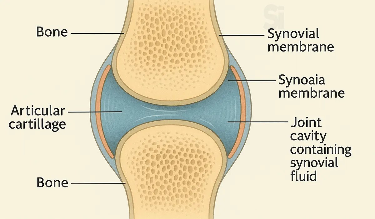
- c. Synovial or freely movable joints
Diagram: Synovial Joint part
Diagram: Synovial Joint part
a. Fibrous or Fixed Joints
Features:
- The bones are connected by fibrous connective tissue.
- There is no joint cavity.
- These joints are immovable.
Examples:
- Sutures of the skull
- Teeth in their sockets (tooth and jaw bone)
b. Cartilaginous or Slightly Movable Joints
Features:
- The bones are joined by cartilage.
- There may be a small gap, but no true joint cavity.
- These joints allow slight movement.
Examples:
- Vertebrae of the spine
- Junction between ribs and sternum
c. Synovial or freely movable joints
It is the most common type of joint found in the human body, and
contains several structures which are not seen in fibrous or
cartilaginous joints.
In this article we shall look at the anatomy of a synovial joint - the
joint capsule, neurovascular structures and clinical correlations.
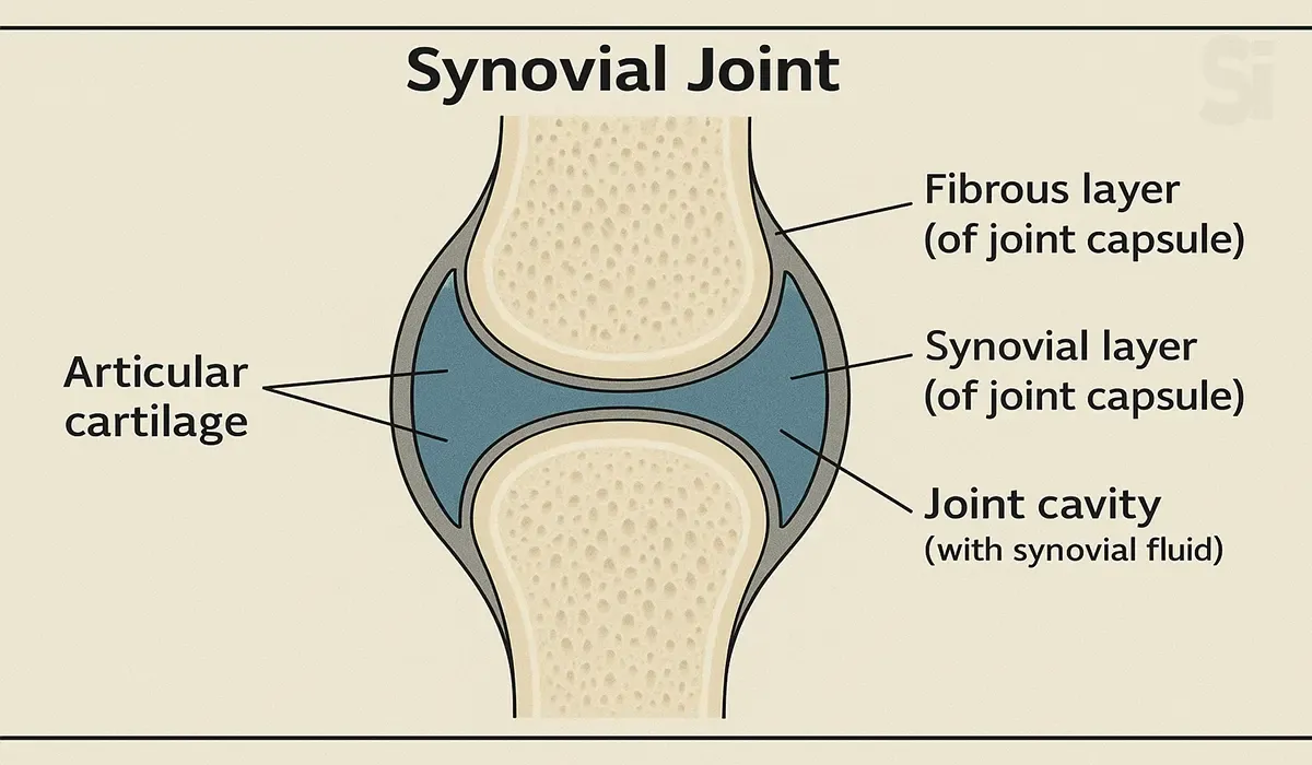
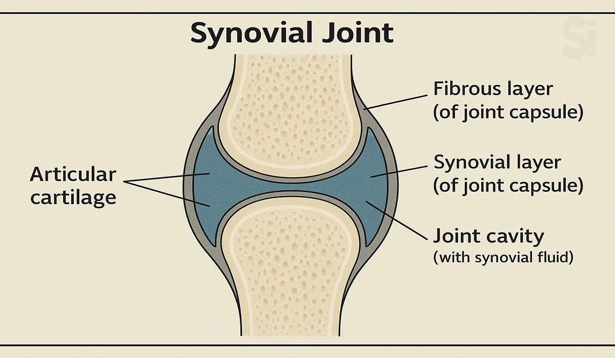
Diagram: Synovial Joint
Key Structures of a Synovial Joint
The three main features of a synovial joint are:
- (i) Articular Capsule
- (ii) Articular Cartilage
- (iii) Synovial Fluid
(i) Articular Capsule: The articular capsule surrounds the joint
and is continuous with the periosteum of articulating bones.
It consists of two layers:
-
Fibrous layer (outer): consists of white fibrous tissue, known
as the capsular ligament. It holds together the articulating bones and
supports the underlying synovium.
-
Synovial layer (inner): a highly vascularized layer of serous
connective tissue. It absorbs and secretes synovial fluid, and is
responsible for the mediation of nutrient exchange between blood and
joint. Also known as the synovium.
(ii) Articular Cartilage: The articulating surfaces of a synovial
joint (i.e. the surfaces that directly contact each other as the bones
move) are covered by a thin layer of hyaline cartilage.
The articular cartilage has two main roles:
- Minimizing friction upon joint movement.
- Absorbing shock.
(iii) Synovial Fluid: The synovial fluid is located within the
joint cavity of a synovial joint.
It has three primary functions:
- Lubrication
- Nutrient distribution
- Shock absorption
Articular cartilage is relatively avascular, and is reliant upon the
passive diffusion of nutrients from the synovial fluid.
Accessory Structures of a Synovial Joint
Accessory Ligaments: The accessory ligaments are separate ligaments
or parts of the joint capsule. They consist of bundles of dense regular
connective tissue, which is highly adapted for resisting strain. This
resists any extreme movements that may damage the joint.
Diagram: Accessory Structures of a Synovial Join
Bursa: A bursa is a small sac lined by synovial membrane,
and filled with synovial fluid.
Bursa are located at key points of friction in a joint. They afford
joints greater freedom of movement, whilst protecting the articular
surfaces from friction-induced degeneration.
Diagram: Bursa
They can become inflamed following infection or irritation by over-use
of the joint (bursitis).
Innervation: Synovial joints have a rich supply from articular
nerves.
The innervation of a joint can be determined using Hilton's Law - 'the
nerves supplying a joint also supply the muscles moving the joint and the
skin covering their distal attachments.
Articular nerves transmit afferent impulses, including proprioceptive
(joint position) and nociceptive (pain) sensation.
Vasculature: Arterial supply to synovial joints is via articular
arteries, which arise from the vessels around the joint. The articular
arteries are located within the joint capsule, mostly in the synovial
membrane.
A common feature of the articular arterial supply is frequent anastomoses
(communications) in order to ensure a blood supply to and across the joint
regardless of its position. In practice this usually means arteries are
above and below a joint, curving round each side of it and joining via
small connecting vessels.
The articular veins accompany the articular arteries and are also found in
the synovial membrane.
Components of a Synovial Joints
- Articular Capsule.
- Synovial Layer (Inner & Outer).
- Articular Cartilage.
- Synovial Fluid.
- Ligament: It is a band of strong muscular tissue.
- Tendon: A cover of strong fibrous connective tissue.
-
Capsule: A two layered membrane that covers the synovial joint.
- Bursae: A pad like sac or cavity close to the joints.
Diagram: Synovial Joints
Synovial Joint: Types
- Gliding joint or Plain joint
- Ball and Socket joint
- Condylar joint
- Pivot joint
- Saddle joint
Diagram: Synovial Joint Types
Properties of Synovial Joint
-
Synovial joints are the most common type of joint in the body.
-
A key structural characteristic for a synovial joint is the
presence of a joint cavity.
-
This synovial fluid filled space is the site at which the
articulating surfaces of the bones contact each other.
-
The articulating bone surfaces at a synovial joint are covered
with fibrous connective tissue or cartilage.
-
This gives the bones of a synovial joint the ability to move
smoothly against each other, allowing for increased joint
mobility.
-
Synovial joints are characterized by the presence of a joint
cavity.
-
The walls of this space are formed by the articular capsule, a
fibrous connective tissue structure that is attached to each bone
just outside the area of the bone's articulating surface.
-
The bones of the joint articulate with each other within the joint
cavity.
- Ligaments are required to bind the bones together
-
At many synovial joints, additional support is provided by the
muscles and their tendons that act across the joint. A tendon is
the dense connective tissue structure that attaches a muscle to
bone.
Muscles: Types
Structure of the body that converts the chemical energy of ATP into
mechanical work.
Diagram: Muscle
Types:
- Striated or Voluntary Muscle : It can move at our will.
-
Visceral or Involuntary muscles : Do not move under our will.
-
Cardiac Muscle : It is also a muscle of involuntary nature.
Functions of Skull, Vertebral Column & Girdles
| Bone |
Function |
|
Skull: Skull is made of mainly brain box (cranium),
which encloses the brain.
|
*Support of the head region.
*Movement of the jaws for articulation and mastication.
*Protection of the brain.
|
|
Vertebrae: Consists of a number of separate bones
called vertebrae. The column is made up of 33 vertebrae.
|
*It supports the axial skeleton.
*Protects and supports the head and spinal cord.
|
|
Girdle Bones: There are two types of girdle bones.
a. Pectoral Girdle & b. Pelvic Girdle
|
*The girdles anchor the limbs to the body in a manner suitable
to their special functions.
|
|
Sternum & Ribs: Sternum consists of a number of
segments called manubrium. Ribs are cage like 12 pairs of
bones.
|
*Protect and delicate organs inside the chest
Carry out breathing movements
|
Pain and Inflammation
Pain: Pain is an unpleasant sensory and emotional experience
associated with acute or potential tissue damage. It is a protective
mechanism for the body.
Inflammation: Inflammation is the active defensive response /
reaction process of tissues against injury, infection etc.
Pain: Types
- Acute pain
- Chronic pain
- Spasmodic pain
Classical signs of Inflammation
- Pain
- Redness
- Heat and
- Swelling
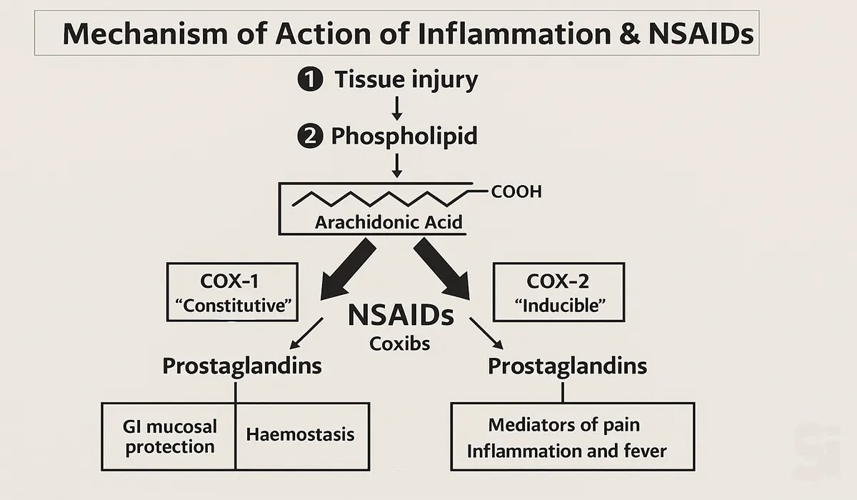
Mechanism of Action of Inflammation & NSAIDs
1. Tissue injury
-
When any part of the body is injured or damaged, the cell membranes
release substances called phospholipids.
2. Formation of arachidonic acid
-
These phospholipids are converted into arachidonic acid, which is
the raw material for producing inflammation-causing chemicals.
3. Prostaglandin production
Arachidonic acid is converted into prostaglandins by two enzymes:
-
COX-1 (“Constitutive”): Always active in the body. It produces
prostaglandins that protect the stomach lining (GI protection) and
help blood clotting (hemostasis).
-
COX-2 (“Inducible”): Produced mainly during injury or inflammation.
It creates prostaglandins that cause pain, swelling, inflammation,
and fever.
4. How NSAIDs work
-
Non-steroidal anti-inflammatory drugs (NSAIDs) like ibuprofen,
naproxen, or diclofenac block both COX-1 and COX-2.
-
By blocking these enzymes, the production of prostaglandins is
reduced, which helps relieve pain, reduce fever, and decrease
inflammation.
5. Why side effects happen
-
Since NSAIDs also block COX-1 (the “good” enzyme), the protective
prostaglandins in the stomach are reduced.
-
This is why long-term NSAID use can cause gastric irritation,
ulcers, or bleeding problems.

Diagram: Mechanism of Action of Inflammation and NSAIDs
👉 In short: When tissue is injured → arachidonic acid forms →
prostaglandins are produced → pain, inflammation, and fever occur.
NSAIDs block this pathway → less prostaglandin → less pain and
inflammation. But because COX-1 is also blocked, side effects like stomach
irritation may happen.
Pain and Inflammation: Synthesis
When the body gets injured or faces infection, inflammatory stimuli
(like trauma, infection, stress) activate special enzymes in the cell
membrane. These enzymes release Arachidonic Acid from the membrane
phospholipids.
Now, Arachidonic Acid can follow two main pathways:
1. Cyclooxygenase (COX) Pathway
COX-1 and COX-2 enzymes convert arachidonic acid into Prostaglandins
and Thromboxanes.
These substances have different effects:
-
Prostaglandins (PGE₂, PGD₂, PGF₂α): cause pain, fever, swelling,
smooth muscle contraction, and protect the stomach lining.
-
Thromboxane A₂ (TXA₂): helps platelets to clump (blood clotting)
and causes blood vessel constriction.
-
Prostacyclin (PGI₂): dilates blood vessels and prevents too much
platelet aggregation.
👉 NSAIDs (like aspirin, ibuprofen) block COX enzymes, reducing
prostaglandins and thromboxanes. This relieves pain, inflammation, and
fever, but may cause side effects like gastric irritation.
2. Lipoxygenase (LOX) Pathway
-
Here, arachidonic acid is converted into Leukotrienes and
Lipoxins.
-
Leukotrienes (LTB₄, LTC₄, LTD₄, LTE₄): increase inflammation,
cause airway constriction (asthma), and attract immune cells.
-
Lipoxins (LXA₄, LXB₄): act as “stop signals” for inflammation,
helping the body to resolve and heal.
Role of Steroids:
-
Steroids (like prednisolone) block the release of arachidonic acid
right at the beginning by inhibiting Phospholipase A₂.
-
This means both COX and LOX pathways are suppressed, giving a
stronger anti-inflammatory effect than NSAIDs.

Diagram: Pain and Inflammation Synthesis1
Diagram: Pain and Inflammation Synthesis2
In Short (Easy Version):
-
Arachidonic Acid = the starting point of pain and inflammation.
-
COX Pathway → Prostaglandins & Thromboxane = pain, fever,
swelling, clotting.
-
LOX Pathway → Leukotrienes = more inflammation, asthma, allergy.
- Lipoxins = reduce inflammation (healing phase).
- NSAIDs stop COX → reduce pain, fever, swelling.
-
Steroids stop everything at the root → stronger effect, but more
side effects.
👉 This combined view explains how pain, fever, swelling, and healing
are controlled inside the body — and how medicines like NSAIDs and
steroids help in controlling them.
Rheumatism: Classification
Systemic disorders of connective tissue, inflammatory arthropathies,
back troubles and soft tissue rheumatism.
Classification:
-
Non
Articular Rheumatism:
Soft tissue involved.
- Articular Rheumatism: Joints involved.
- Others: Infective.
1) Non Articular Rheumatism
Tendinitis:
The Inflammation of tendon sheath, characterized by local tenderness
around the joint.
Bursitis:
Inflammation of bursa. Often occurs in shoulders and knee joints.
Capsulitis:
Inflammation of joint capsule, Particularly of a shoulder joint. It is
also called Frozen shoulder.
Fibrositis:
Inflammation of muscle characterized by localized pain and stiffness.
Often observed in the neck, shoulders, chest or back.
Epicondylitis:
Inflammation of epicondyle (a rounded bone projected from the
articular end of a bone).
2) Articular Rheumatism
Osteoarthritis: It is a non-inflammatory joint disease
characterized by the degeneration of articular cartilage.
Rheumatoid Arthritis: It is a most common and chronic form of
arthritis. It involves more than one joint.
Articular Rheumatism : Sign and Symptoms:
- Starts gradually.
- Pain and stiffness is common.
- Patients complain of early morning stiffness.
-
Progression of the disease with marked inflammation is observed.
- Tests of rheumatoid factors are positive in 70-80% of patients.
The word juvenile means childhood. Usually it occurs in children under 16
years of age. It is characterized by joint inflammation along with
high-grade fever.
Articular Rheumatism: Ankylosing Spondylitis
Ankylosing Spondylitis is a progressive chronic arthritis. The word
Ankylosis means stiff joint condition and Spondylitis means inflammation
of vertebrae.
Sign and symptoms:
- Pain is worse after exercise.
- Low back pain and morning stiffness.
- Pain around the ribs may.
- Marked rigidity of spine may occur.
3) Other types of Rheumatism
Septic Arthritis: Inflammation of joints caused by pus
producing microorganisms. Usually Staphylococcus, Streptococcus is the
causative organism.
Osteomyelitis:
Inflammation of bone marrow caused by pus producing pathogens.
Other Painful Conditions
Sprain:
Injury to ligaments that causes pain and loss of movement.
Strain:
Trauma to the muscle that results from excessive physical effort.
Arthralgia:
Pain within a joint without any definite cause of joint disease.
Dysmenorrhoea: Painful or difficulty in menstruation.
Sciatica:
Severe pain felt at the back of the thigh.
Lumbago:
Slow but continuous pain in the lumbar part of the back.
Dislocation:
Displacement of bone from its normal location.
Renal colic: Colic means pain, resulting from periodic spasm in an
abdominal organ. Renal colic means spasm of ureter due to a stone.
Gout:
Painful condition due to deposition of uric acid crystal in the small
peripheral joints and the tissues around.
Sign and Symptoms of Gout:
-
Sudden onset of severe pain with marked inflammation and tenderness.
- Fever, sweating, Loss of appetite.
- Raised serum uric acid level.
Clinical Relevance: Osteoarthritis
Osteoarthritis is the most common form of joint inflammation
(arthritis). It stems from heavy use of articular joints over the
course of many years, which can result in the wearing away of
articular cartilage, and often the erosion of the underlying
articulating surfaces of bones as well.
The changes which occur are irreversible and degenerative. This
results in the decreased effectiveness of articular cartilage as a
shock absorber and lubricated surface, as well as the roughened
edges causing further damage.
As a result of this degeneration, repeated friction can cause
symptoms of joint pain, stiffness and discomfort. This condition
usually affects joints that support full body weight, such as the
hips and the knees.
Diagram: Clinical Relevance Osteoarthritis
Arthritis can also come about through other causes, including;
-
As a result of infection, due to the ease with which blood (and
any associated bacteria) can enter the joint cavity via the
synovial membrane;
-
Due to auto-inflammatory causes, as in rheumatoid arthritis, or;
-
As a result of infection but not involving infection of the
joint itself, as in reactive arthritis.
FAQs
1. What is the full form of RA?
The full form of RA is Rheumatoid Arthritis.
2. What is the full form of JRA?
The full form of JRA is Juvenile Rheumatoid Arthritis.
3. What is the musculoskeletal system in simple words?
The musculoskeletal system is the network of bones, muscles, and
joints in your body. It allows you to move, stand, walk, and perform
daily tasks, while also giving structure and support to the entire
body.
4. Why is the musculoskeletal system important for Pharmaceutical
Sales Representatives to understand?
As a Pharmaceutical Sales Representative, having a clear understanding
of this system makes it easier to explain how certain medications
work, especially drugs related to bone and muscle health. It also
helps you communicate more confidently with healthcare professionals.
5. How does the musculoskeletal system work together?
Bones act as the framework, muscles provide movement, and joints
connect the parts. Together, they work like a well-coordinated team,
allowing the body to move smoothly and stay strong.
6. What problems can affect the musculoskeletal system?
Common problems include arthritis, back pain, muscle strain,
osteoporosis, and bone fractures. A strong knowledge of these
conditions helps a Pharmaceutical Sales Representative explain
treatment options more effectively to doctors.
Conclusion
For anyone starting a career as a Pharmaceutical Sales Representative
(PSR) or Medical Representative (MR), understanding The Human
Musculoskeletal System and Its Functions is essential. This knowledge
not only gives you the confidence to discuss bone and muscle-related
medicines with doctors, but it also helps you build credibility as a
professional. Always remember, mastering the basics is the first step
to long-term success in pharmaceutical sales. Keep learning, keep
practicing, and stay curious!