
The Human Digestive System and Its Functions
Want to become a successful Pharmaceutical Sales Representative? This
article will guide you through The Human Digestive System and Its
Functions in a clear, step-by-step way. By exploring each part of the
system, you’ll gain practical knowledge that can help you better
understand the products you represent and communicate more effectively
with healthcare professionals.

Diagram: Human digestive system
This resource is designed for new or aspiring Medical Sales
Representatives, or anyone preparing to build a career in the
pharmaceutical industry. It’s written in a simple and approachable style,
keeping the content easy to follow and relevant to your future role.
If you’re planning to join the medical sales field, this guide is a great
place to start. Think of it as your first step toward developing the
knowledge and confidence you’ll need for a rewarding career as a Sales
Representative.
Table of Contents: The Human Digestive System and Its Functions
Table of Contents of this Article-
- The Human Digestive System and Its Functions
- The Components of Digestive System
- Esophagus
- Stomach
- Intestine
- Accessory Organs of Digestive System
- Digestive Juices
- Saliva
- Composition of Gastric Juice
- Pepsin
- Mucus
- Function of Hydrochloric Acid
- What is Prostaglandins?
- Types of Factors in GIT
- FAQs
- Conclusion
The Human Digestive System and Its Functions
The human digestive system is responsible for breaking down food so the
body can absorb essential nutrients. It involves several key organs,
including the mouth, esophagus, stomach, and the small and large
intestines.
Functions of the Human Digestive System
Step-by-Step functions 👇
1. Ingestion — taking food in
- Food enters the mouth, where teeth break it into smaller pieces and the tongue helps position and mix it.
- Saliva moistens the food, starts carbohydrate digestion (salivary amylase), and helps form the bolus for swallowing.
2. Swallowing and propulsion — moving food along
- Swallowing has a voluntary oral phase and then automatic phases that close off the airway while the bolus moves into the esophagus.
- The esophagus uses coordinated muscle contractions called peristalsis to push the bolus toward the stomach.
- Two sphincters—upper esophageal sphincter (UES) and lower esophageal sphincter (LES)—open and close to control passage and prevent backflow.
3. Mechanical digestion & stomach processing — mixing and breaking down
- The stomach stores food and uses strong muscular contractions to churn and mechanically break the bolus into smaller particles.
- Stomach secretions (HCl, pepsinogen → pepsin, and mucus) perform chemical digestion, especially of proteins, and create an acidic environment that kills many microbes.
- The stomach controls how quickly small amounts of semi-digested food (chyme) are released into the small intestine—this is gastric emptying.
4. Chemical digestion in the small intestine — finishing the breakdown
- The duodenum is where most chemical digestion is completed: pancreatic enzymes (amylase, lipase, proteases) and bile from the liver/gallbladder act on chyme.
- Pancreatic bicarbonate neutralizes stomach acid so intestinal enzymes can work.
- The small intestine’s inner surface (vill i and microvilli) increases surface area for digestion and absorption.
5. Absorption — moving nutrients into the body
- Carbohydrates → broken into monosaccharides (e.g., glucose) and absorbed into the bloodstream via transporters.
- Proteins → broken into amino acids and small peptides, absorbed via active transport.
- Fats → emulsified by bile into tiny droplets, digested by lipase into fatty acids and monoglycerides, assembled into chylomicrons inside enterocytes, and transported into the lymphatic system (lacteals).
- Vitamins & minerals: water-soluble vitamins (B, C) are absorbed directly; fat-soluble vitamins (A, D, E, K) need fats and bile; minerals like iron require specific forms (ferrous) and transport systems.
- Water is absorbed throughout the small intestine and further in the colon.
6. Large intestine — reclaiming water and forming stool
- The colon absorbs remaining water and electrolytes, concentrating waste into stool.
- Gut bacteria ferment undigested fibers to produce short-chain fatty acids (useful energy source) and synthesize certain vitamins (e.g., vitamin K).
- The rectum stores stool until defecation, controlled by internal and external anal sphincters.
7. Regulation, coordination & protection
- Digestion is coordinated by the nervous system (enteric nervous system + autonomic input) and hormones (gastrin, secretin, cholecystokinin).
- Protective mechanisms include mucus, bicarbonate, the acidic gastric environment, and the mucosal immune system (plus the microbiome). These keep pathogens in check and protect the lining.
Knowing where digestion happens and which secretions act where helps you
explain how drugs work (e.g., antacids/PPIs reduce HCl, pancreatic
enzyme replacements aid malabsorption, bile acid drugs affect fat
absorption).
A clear, simple explanation of these steps makes your conversations with
clinicians more credible and focused on mechanism and patient benefit.
The Components of Digestive System
| Gastro Intestinal Tract | Accessories Organs |
|---|---|
| 1. Oral Cavity | 1. Liver |
| 2. Salivary glands | 2. Pancreases |
| 3. Pharynx | 3. Gall Bladder |
| 4. Esophagus | 4. Salivary glads |
| 5. Stomach | |
| 6. Small Intestine | |
| 7. Large Intestine | |
| 8. Rectum | |
| 9. Anus |
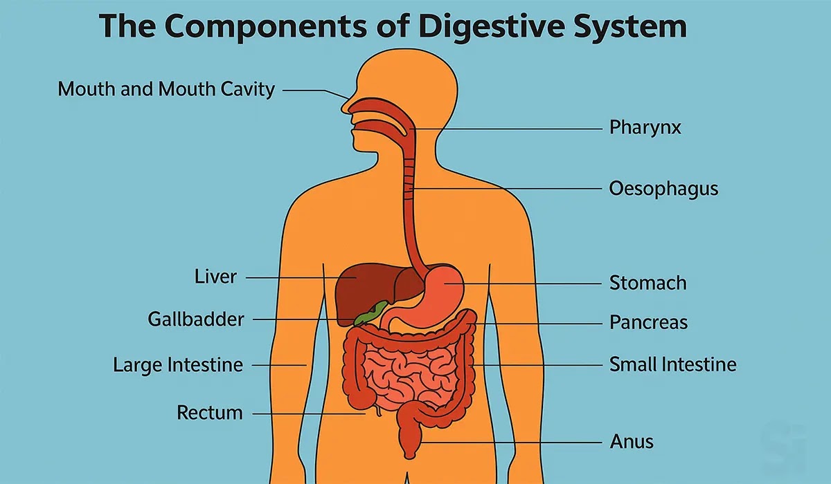
Diagram: The Components of Digestive System
Gastro Intestinal Tract
Different Parts:
*Mouth / Oral Cavity
- Lip
- Teeth
- Tongue
*Pharynx
*Esophagus
*Stomach
- Cardia
- Fundus
- Body
- Pylorus
*Small Intestine
- Duodenum
- Jejunum
- Ileum
*Large intestine
- Caecum
- Ascending colon
- Transverse colon
- Descending colon
- Sigmoid colon
*Rectum
*Anus
Accessory Organ
Different Parts:
*Salivary Gland
- Parotid (below the ear)
- Submandibular (below the jaw)
- Sublingual (below the tongue)
*Liver
*Pancreas
*Gallbladder

Diagram: Alimentary Tract and Accessory Organ
Esophagus
The esophagus is a muscular tube that connects the throat (pharynx) to
the stomach. It is about 25 centimeters (10 inches) long in adults and
runs behind the trachea and heart, eventually passing through the
diaphragm before joining the stomach.
Food does not simply “fall” through the esophagus. Instead, the walls
of the esophagus contain smooth muscles that contract in a
coordinated, wave-like motion known as peristalsis. This action pushes
the swallowed food (called a bolus) steadily down toward the stomach.
At the lower end, the esophagus is connected to the stomach through a
muscular ring called the lower esophageal sphincter (LES). This
sphincter acts like a valve or gatekeeper—it opens to allow food to
enter the stomach and then closes tightly to prevent stomach acid and
food from flowing back up. If this sphincter becomes weak or relaxed,
it can lead to problems like acid reflux (GERD).
Function:
- Transports food and liquids from the pharynx to the stomach.
- Uses peristaltic movements to ensure smooth passage of the food bolus.
- The lower esophageal sphincter prevents backflow of acidic contents into the esophagus, protecting it from damage.
Stomach
The stomach is a hollow muscular bag situated between the
esophagus and small intestine.

Diagram: Stomach
Functions:
- Storage of Food
- esophagus
- Mixing of Food
- Controlling of food movements
- Digestion
Parts of Stomach
It is histologically divided into 4 portions:
- Cardia (Upper part): Connects the esophagus and the stomach.
- Fundus: Projects upward in a dome shaped manner.
- Corpus: Major part of the stomach.
- Pylorus (Lower part): Contains the pyloric sphincter.

Diagram: Parts of Stomach
Stomach: Musculature
- a. Mucosa
- b. Submucosa
- c. Muscular Layer
- d. Serosa

Diagram: Stomach Musculature
Stomach: Glands
Gland of Gastric Walls:
- a. Gastric gland
- b. Pyloric gland

Diagram: Stomach Glands
Gastric Gland
- Mucous Neck Cells: Localized to the neck region, that secrete a mucous.
- Peptic / Chief Cells: Located deep within the glands secretes pepsinogen.
- Parietal Oxyntic cells: Mostly in the middle and upper portions of the gland, with an intracellular canalicular system, secreting HCI and Intrinsic Factor.

Diagram: Gastric Gland
Pyloric Gland
- Mucous Neck Cells (Mostly): Localized to the neck region, that secrete mucus.
- Peptic / Chief Cells (Few): Located deep within the glands secretes pepsinogen.
- Gastrin Cells: Secretes gastrin, a local hormone responsible for Hydrochloric acid secretion.
Intestine
The intestines are divided into two main parts: the
small intestine and the large intestine.
Small Intestine
It is subdivided into three parts:
- Duodenum
- Jejunum
- Neum
Small Intestine has finger-like projections known as villi,
where absorption takes place.

Diagram: Small Intestine
Function:
- Major site of digestion and absorption of nutrients.
- Site of secretion of salts.
Large Intestine
The large intestine has following 5 sections:
- Caecum
- Ascending colon
- Transverse colon
- Descending colon
- Sigmoid colon
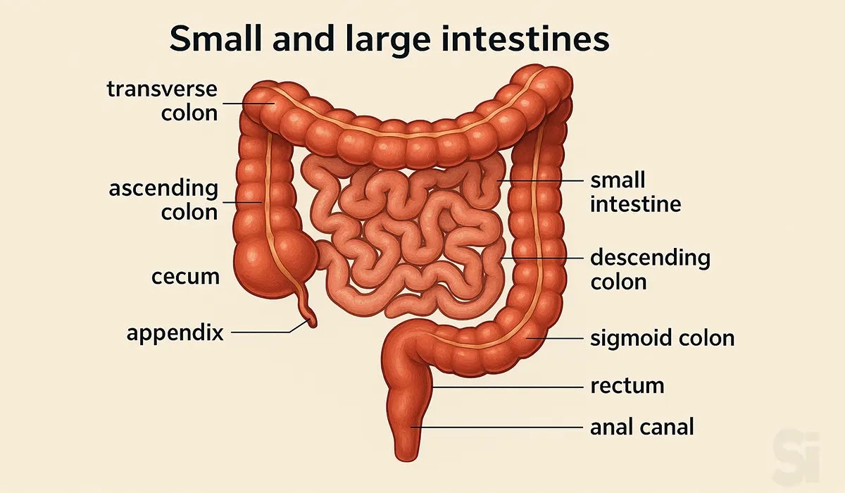
Diagram: Small and large intestine
Accessory Organs of Digestive System
The digestive system is not limited to the stomach and
intestines alone. There are also several accessory
organs that play a vital supporting role in digestion.
These organs do not directly handle the food, but they
produce and release important substances that help the
body break down nutrients effectively.
1. Salivary Glands
The salivary glands are located in and around the
mouth. They produce saliva, which contains enzymes
like amylase that start breaking down carbohydrates
while food is still in the mouth. Saliva also moistens
food, making it easier to chew and swallow.
2. Pancreas
The pancreas lies behind the stomach and has two major
roles. First, it produces digestive enzymes (lipase,
amylase, protease) that help break down fats,
carbohydrates, and proteins in the small intestine.
Second, it releases bicarbonate to neutralize stomach
acid as food enters the intestine. The pancreas also
has an endocrine role, producing hormones like
insulin, which regulates blood sugar.
3. Liver
The liver is the largest internal organ and acts as
the body’s chemical factory. Its main digestive
function is producing bile, a fluid that helps break
down and absorb dietary fats. The liver also processes
nutrients absorbed from the small intestine,
detoxifies harmful substances, and stores vitamins and
minerals for future use.
4. Gallbladder
The gallbladder is a small pouch located beneath the
liver. Its main role is to store and release bile when
fatty food enters the small intestine. By squeezing
bile into the intestine at the right time, the
gallbladder makes fat digestion more efficient.

Diagram: Accessory Organs of Digestive System
Digestive Juices
For food to be broken down into nutrients the body can
use, the digestive system relies on a variety of
digestive juices. These fluids are secreted by
different organs at different stages of the process.
Each type of juice contains special enzymes or
substances that target specific nutrients like
carbohydrates, proteins, and fats.
1. Saliva
Produced by the salivary glands in the mouth, saliva
contains the enzyme amylase, which begins breaking
down starch into simple sugars. Saliva also moistens
food, making it easier to chew, swallow, and move
smoothly down the esophagus.
2. Gastric Juices
Secreted by the stomach lining, gastric juice is a
mixture of hydrochloric acid (HCl), pepsinogen, and
mucus. HCl creates an acidic environment, which
activates pepsin to digest proteins, while mucus
protects the stomach lining from damage.
3. Pancreatic Juices
The pancreas produces powerful digestive enzymes,
including lipase (for fats), amylase (for
carbohydrates), and trypsin (for proteins). These
enzymes are released into the small intestine, along
with bicarbonate, which neutralizes stomach acid and
allows enzymes to work effectively.
4. Intestinal Juices
The small intestine secretes its own juices, often
called succus entericus, which contain enzymes that
complete the final steps of digestion. For example,
maltase, lactase, and sucrase break down sugars into
glucose and other simple forms that the body can
absorb.
5. Bile
Bile is produced by the liver and stored in the
gallbladder. It doesn’t contain enzymes but has bile
salts, which help break large fat globules into
smaller droplets—a process called emulsification. This
makes it easier for pancreatic lipase to digest fats
efficiently.

Diagram: Digestive Juice
Types of Secretion
| Types of Secretion | Daily Volume (mi) | pH |
|---|---|---|
| Saliva | 1000 | 6.0-7.0 |
| Gastric secretion | 1500 | 1.0-3.5 |
| Pancreatic secretion | 1000 | 8.0-8.3 |
| Bile | 1000 | 7.8 |
| Small intestine secretion | 1800 | 7.5-8.0 |
| Brunner's gland secretion | 200 | 8.0-8.9 |
| Large intestinal secretion | 200 | 7.5-8.0 |
| Total | 6700 |
Saliva
Saliva is the first digestive fluid that comes into
play as soon as food enters the mouth. Produced by the
salivary glands, it performs several important
functions that prepare food for its journey through
the digestive system.
Functions of Saliva
Moistening and lubrication: Saliva softens and
moistens food, forming it into a bolus that can be
swallowed easily without damaging the lining of the
throat or esophagus.
Taste perception: Many substances must first
dissolve in saliva before the taste buds can detect
them, making saliva essential for the sense of taste.
Starting digestion: Saliva contains important
enzymes:
- Amylase begins breaking down carbohydrates into simpler sugars.
- Lingual lipase starts the digestion of fats, especially in infants.
Speech and comfort: By keeping the mouth moist,
saliva helps in clear speech and prevents dryness or
discomfort.
Protective role: Saliva also contains
antibacterial compounds and maintains oral hygiene,
reducing the risk of infections.

Composition of Gastric Juice
- Pepsin : Active form precursor pepsinogen.
- Hydrochloric acid (HCl) : Activates pepsinogen.
- Mucus : Provides mechanical protection for gastric mucosa.
- Intrinsic factor : Essential to the absorption of Vitamin B12.
- Gastrin : Hormone that stimulates HCL secretion.

Pepsin
Pepsin is one of the most important enzymes in the
stomach, responsible for beginning the digestion of
proteins. It is secreted by the stomach lining in an
inactive form called pepsinogen.
When food enters the stomach, hydrochloric acid (HCl)
is released. This acidic environment converts
pepsinogen into its active form, pepsin. Once active,
pepsin starts breaking down large protein molecules
into smaller fragments known as peptones.
This is only a partial digestion step—later enzymes in
the small intestine will continue the process to
release amino acids that the body can absorb.
Equation Summary:
- Pepsinogen (inactive) + HCl → Pepsin (active)
- Protein + Pepsin → Peptones (partially digested proteins)

Mucus
Mucus is a vital protective secretion in the stomach
and throughout the gastrointestinal tract. It is
mainly made up of water, glycoproteins, and
bicarbonate ions, which together create a protective
barrier between the stomach lining and the harsh
acidic environment inside.
Functions of Mucus
a. Chemical Protection
- The bicarbonate in mucus reacts with hydrochloric acid (HCl), creating a neutralizing effect.
- This helps maintain a more balanced pH at the surface of the stomach lining, preventing acid-related injury.
b. Mechanical Protection (Gastric Mucosal
Barrier)
- Mucus forms a thin but strong coating over the gastric mucosa, known as the gastric mucosal barrier.
- This layer protects the delicate stomach lining from being digested or damaged by acid (HCl) and pepsin.
- When this barrier is weakened—for example, by NSAIDs, alcohol, or Helicobacter pylori infection—it can lead to ulcers and other gastric problems.
c. Lubrication
- Mucus keeps the stomach lining moist and slippery, allowing food to move smoothly during digestion without friction or irritation.
Function of Hydrochloric Acid
Hydrochloric acid (HCl) is one of the most important
components of gastric juice, and it plays multiple
roles in digestion and overall gut health. Without
HCl, the stomach would not be able to properly break
down food or protect the body from harmful microbes.
Its functions can be grouped into three main
categories:
A. Digestion and Absorption
Activation of pepsin: HCl converts inactive
pepsinogen into pepsin, the active enzyme that
digests proteins.
Breakdown of food substances: It helps in the
hydrolysis of proteins, allowing large molecules to
be split into smaller peptides for easier digestion.
Iron absorption: HCl keeps dietary iron in its
ferrous (Fe²⁺) state, which is the form that can be
absorbed in the small intestine. This is essential
for preventing iron deficiency and anemia.
B. Bactericidal Action
HCl creates a highly acidic environment (pH 1–2),
which kills many harmful bacteria and pathogens that
may enter the body through food and water.
This protective action reduces the risk of
infections in the gastrointestinal tract.
C. Creating an Optimal Environment
The acidic pH maintained by HCl provides the ideal
environment for enzymes like pepsin to function
efficiently.
It also ensures that food is properly broken down
before it enters the small intestine for nutrient
absorption.
Hydrochloric Acid Secretion: Mechanism
In the stomach, special cells called parietal cells
are responsible for producing hydrochloric acid (HCl).
This acid is essential because it helps break down
food, activates digestive enzymes, and protects the
body by killing harmful bacteria.
The process starts when signals from the body—such as
acetylcholine, histamine, and gastrin—tell the
parietal cells to start working. Inside the cell,
water (H₂O) and carbon dioxide (CO₂) combine to form
carbonic acid, which quickly splits into two parts:
bicarbonate (HCO₃⁻) and hydrogen ions (H⁺).
The hydrogen ions are pumped out of the cell into the
stomach cavity through a special system called the
proton pump (H⁺/K⁺ ATPase pump). At the same time,
chloride ions (Cl⁻) move out of the cell and join the
hydrogen ions. Together, they form HCl, or
hydrochloric acid.
Meanwhile, the bicarbonate formed inside the cell is
exchanged with chloride ions from the blood, which
keeps the whole process balanced.

Diagram: Hydrochloric Acid Secretion
Mechanism

Diagram: Hydrochloric Acid Secretion Mechanism
In Short-
- Parietal cells make hydrochloric acid.
- Signals (acetylcholine, histamine, gastrin) activate the cells.
- Hydrogen and chloride ions combine in the stomach to form HCl.
- The proton pump is the key player in moving hydrogen out of the cell.
This step-by-step mechanism ensures the stomach has
the acid it needs to digest food efficiently.
Hydrochloric Acid Production in Parietal Cell: Mechanism
Inside the stomach lining, parietal cells are the
main factories for hydrochloric acid (HCl). The
process begins when carbon dioxide (CO₂) from the
blood combines with water (H₂O) inside the cell.
With the help of an enzyme called carbonic
anhydrase, this mixture forms carbonic acid
(H₂CO₃).
Carbonic acid quickly breaks down into bicarbonate
(HCO₃⁻) and hydrogen ions (H⁺).
- The hydrogen ions (H⁺) are pumped out into the stomach cavity through a special system known as the H⁺/K⁺ ATPase pump, often called the proton pump. This pump uses energy (ATP) to exchange hydrogen ions from the cell with potassium ions (K⁺) from the stomach.
- At the same time, bicarbonate (HCO₃⁻) moves out of the parietal cell into the blood. In exchange, chloride ions (Cl⁻) come into the cell and then pass into the stomach cavity.
Finally, when hydrogen ions (H⁺) meet chloride
ions (Cl⁻) in the stomach, they form hydrochloric
acid (HCl).

Diagram: Hydrochloric Acid Production in
Parietal Cell Mechanism
Quick Summary-
- CO₂ + H₂O → forms carbonic acid (H₂CO₃).
- Carbonic acid splits into bicarbonate (HCO₃⁻) and hydrogen (H⁺).
- Hydrogen (H⁺) pumped out by proton pump, chloride (Cl⁻) enters the stomach.
- H⁺ + Cl⁻ = Hydrochloric Acid (HCl).
This is how your stomach produces the strong acid
needed to break down food and protect against
harmful microbes.
Hydrochloric Acid Production
Acid production in parietal cell is stimulated
by:
- Acetylcholine
- Histamine
- Gastrin
How They Work:
- A. Acetylcholine + M2 Receptor = ↑ Acid Production + Histamine Secretion
- B. Histamine + H2 Receptor = ↑ Gastric Acid Secretion
- C. Gastrin + Gastrin Receptor = ↑ HCL Secretion
What is Prostaglandins?
Prostaglandins are natural chemical messengers
made from fatty acids in the body. They are not
hormones, but they act very much like hormones
because they send signals that help regulate
different functions in many organs. In the
stomach, prostaglandins play a protective role.
One of the most important types is Prostaglandin
E (PGE). This compound helps the stomach stay
healthy in several ways:
1. Stimulates Mucus and Bicarbonate
Secretion:
PGE encourages the stomach lining to release
more mucus and bicarbonate.
- Mucus forms a protective coating on the stomach wall.
- Bicarbonate neutralizes excess acid by balancing pH levels near the stomach lining.
- Together, these actions protect the cells of the stomach from being damaged by hydrochloric acid and digestive enzymes.
2. Supports Blood Flow:
PGE ensures a steady supply of oxygen and
nutrients to the stomach lining by improving
blood circulation. This helps the cells stay
healthy and repair themselves quickly if they
get injured.
3. Maintains Gastric Cell Function:
With enough oxygen, nutrients, and bicarbonate,
gastric cells can work efficiently. They keep
producing protective substances and can resist
the harmful effects of excess acid, alcohol,
NSAIDs, or infections like H. pylori.
Types of Factors in GIT
The gastrointestinal tract (GIT) is constantly
exposed to different substances that can either
protect it or damage it. On one side, there are
aggressive factors such as gastric acid, digestive
enzymes, harmful bacteria, or even external agents
like alcohol and certain medications.
These elements can break down the protective
lining of the stomach and intestines if they
become too strong. On the other side, the body has
defensive factors like mucus, bicarbonate, and
epithelial cells that form a barrier and keep the
stomach safe from injury.
The balance between these aggressive and defensive
factors is what maintains a healthy digestive
system, and when that balance is disturbed,
problems such as gastritis, ulcers, or reflux can
occur.
Aggressive Factors
1. Gastric Juice Barrier:
- Acid
- Pepsin
- Bile salts
- Pancreatic enzymes
2. Endogenous Bacteria:
3. Extrinsic Factor:
- Aspirin
- NSAIDS
- Alcohol
- Caffeine
Defensive Factors
- 1. Mechanical: Mucus
- 2. Chemical Barriers: Bicarbonate
- 3. Biological Barriers: Epithelial cell
FAQs
1) What is the full form of GERD?
Answer: GERD stands for Gastroesophageal
Reflux Disease.
2) What is the full form of HCl?
Answer: HCl stands for hydrochloric acid.
3) What is the full form of GIT?
Answer: GIT stands for Gastrointestinal Tract.
4) What are the main organs of the human
digestive system?
Answer: The main organs are the mouth,
esophagus, stomach, small intestine, large
intestine, liver, pancreas, and gallbladder.
Each organ has a specific role in breaking
down food and absorbing nutrients.
5) Why is the digestive system important
for overall health?
Answer: The digestive system converts the food
we eat into the nutrients the body needs for
energy, growth, and repair. Healthy digestion
helps prevent common problems such as gas,
bloating, constipation, and nutrient
deficiencies, and it also supports immune
function and steady energy levels.
6) How can I keep my digestive system
healthy naturally?
Answer: Eat a balanced, fiber-rich diet, drink
plenty of water, stay active, and avoid
overeating. Probiotics (from yogurt and
fermented foods) can help maintain a healthy
gut microbiome. Also cut down on processed
foods, limit alcohol and caffeine, avoid
smoking, and manage stress.
7) What are the most common digestive
disorders that Medical Sales Representatives
should know about?
Answer: Important conditions to know include
acid reflux (GERD), peptic ulcers, irritable
bowel syndrome (IBS), constipation, diarrhea,
and gastrointestinal infections. Understanding
these helps when discussing relevant
medications and treatment approaches with
healthcare professionals.
8) How do digestive enzymes and stomach
acids function in digestion?
Answer: Digestive enzymes break carbohydrates,
proteins, and fats into smaller units the body
can absorb. Stomach acid (mainly HCl) helps
activate enzymes like pepsin and helps kill
many harmful microbes. A proper balance
between enzymes and acid is essential for
efficient digestion.
Conclusion
Understanding The Human Digestive System and
Its Functions is a key step for anyone
preparing to work in pharmaceutical or medical
sales. A solid grasp of basic anatomy and
physiology lets you discuss mechanisms of
action clearly, build trust with clinicians,
and position products more effectively. Use
this guide as a foundation: keep learning,
stay curious, and practice explaining these
concepts simply — that clarity will make you a
stronger, more credible Sales Representative.

Sarwar Hossain ✅
I am a travel enthusiast and sports-loving person. Education: BBA & MBA in Accounting.