
The Nervous System and Its Functions
This article explains The Nervous System and Its Functions in a clear and
straightforward way, making it easy for beginners to follow along. If you are
preparing to work as a Pharmaceutical Sales Representative—or even as a
Medical Representative—this guide will help you understand the essential
concepts that are often discussed in the medical field.

Diagram: Nervous system diagram
You will get a step-by-step idea of how the human nervous system functions and
why it plays such a central role in our body. The content is simple,
beginner-friendly, and designed to help you build a solid foundation in
medical science.
Table of contents: The Nervous System and Its Functions
Take a look at everything you can learn from this article.
The Nervous System and Its Functions
The Nervous System and Its Functions includes the brain, spinal cord, and
nerves, which work together to control the body’s actions and responses.
With this guide, you can easily understand how messages travel through the
nerves and how the system coordinates different body functions.
This explanation is especially useful if you are preparing for a career as a
Pharmaceutical Sales Representative, because having a clear understanding of
basic medical science gives you a strong advantage in the field.
Now, let’s dive into a detailed discussion about the nervous system....
Nervous System: The nervous system is the chief controlling and
coordinating system of the body.

This system is responsible for providing conscious or unconscious control
of-
- Basic motor and sensory activity.
- Emotional and intellectual function.
Classification of Nervous System
The nervous system is broadly divided into two major parts:
1. Central Nervous System (CNS)
The CNS acts as the main control center of the body.
Brain:
- Receives and processes sensory information.
- Initiates responses and controls thoughts and emotions.
Spinal Cord:
- Conducts signals to and from the brain.
- Controls reflex activities that allow quick responses without brain involvement.
2. Peripheral Nervous System (PNS)
The PNS connects the CNS to the rest of the body, including muscles,
glands, and sensory organs. It has two main parts:
(a) Sensory Neurons
- Carry information from sensory organs (eyes, ears, skin, etc.) to the CNS.
(b) Motor Neurons
- Transmit signals from the CNS to muscles and glands.
- Divided into two systems:
1. Somatic Nervous System
- Controls voluntary movements (like walking, writing, speaking).
2. Autonomic Nervous System
(ANS)
- Controls involuntary activities (like heartbeat, breathing, digestion).
- The ANS itself has two divisions:
Sympathetic Division → Prepares
the body for “fight or flight” during stress or emergency.
Parasympathetic Division → Helps the
body relax and perform “rest and digest” functions after stress.

Classification of Nervous System in details
The nervous system is like the body’s main communication network, and
it can be divided into two big parts: the Central Nervous System (CNS)
and the Peripheral Nervous System (PNS).
The CNS is the control center. It has two key players:
- The brain, which processes information, makes decisions, and shapes our thoughts and emotions.
- The spinal cord, which acts like a highway, carrying messages back and forth between the brain and the rest of the body. It also handles quick reflexes without even asking the brain for help.
The PNS connects the CNS to the rest of the body. Think of it as the
network of wires that keep everything linked together. It has two main
types of nerves:
- Sensory nerves, which bring information from your senses—like touch, sight, and sound—straight to the CNS.
- Motor nerves, which carry instructions from the CNS to your muscles and glands so you can move or react.
Motor nerves are further split into two systems:
- Somatic Nervous System – this controls voluntary actions, like walking, talking, or writing.
- Autonomic Nervous System (ANS) – this runs the things you don’t have to think about, like your heartbeat, digestion, and breathing.
The ANS itself has two sides working in balance:
- The Sympathetic Division, which kicks in when you’re stressed or in danger, preparing your body for a “fight or flight” response.
- The Parasympathetic Division, which does the opposite—it calms you down, helping your body rest, recover, and digest food.

Central Nervous System
The Central Nervous System is the command center of the body. It has
two main parts: the brain and the spinal cord. Together, they
control almost everything we do—from our movements to our emotions.
1. Brain
The brain is the most complex organ, and it is divided into
different parts, each with a specific role:
- Cerebrum: The largest part of the brain, responsible for thinking, memory, reasoning, learning, and voluntary actions.
- Corpus Callosum: A bridge that connects the left and right hemispheres of the brain so they can share information.
- Thalamus: Works like a relay station—it receives sensory information and directs it to the right areas of the brain.
- Hypothalamus: Controls basic functions like hunger, thirst, body temperature, and even emotions. It also links the nervous system with the endocrine system.
- Cerebellum: Maintains balance, posture, and coordination of movements.
- Pons: Connects different parts of the brain and helps regulate breathing.
- Medulla Oblongata: Controls vital involuntary functions such as heartbeat, breathing, and blood pressure.
- Temporal Lobe: Involved in processing sounds, understanding language, and forming memories.
2. Spinal Cord
The spinal cord is a long bundle of nerves running down the back. It
acts as a communication highway between the brain and the body.
It is divided into regions:
- Cervical region (C1–C8): Controls the neck, diaphragm, shoulders, arms, and fingers.
- Thoracic region (T1–T12): Handles chest muscles, abdominal muscles, and some reflex activities.
- Lumbar region (L1–L5): Controls hips, legs, and lower body functions.
- Sacral region (S1–S5): Linked to the bladder, bowel, sexual functions, and lower limbs.
- Coccygeal region: The very end of the spinal cord, with limited but important roles in pelvic functions.

Diagram: Central nervous system diagram 1
If you look at the diagram of the spinal cord, you’ll notice that each
section is linked to specific parts of the body. For example, the
cervical region (at the top) controls the neck muscles, shoulders, and
even your fingers.
Moving down, the thoracic region connects with the chest and abdominal
muscles, while the lumbar region manages the hips and legs. The sacral
nerves handle very important functions like bladder control, bowel
movements, and sexual functions. Finally, the coccygeal region, at the
very bottom, plays a role in pelvic functions.
In the brain diagram, each labeled part has a unique role. The cerebrum takes care of thinking and voluntary actions, the cerebellum helps you keep balance, and the medulla oblongata keeps your heart beating and lungs working without you even noticing. Together, these structures make sure your body runs smoothly, whether you’re walking, thinking, or simply breathing.


In the brain diagram, each labeled part has a unique role. The cerebrum takes care of thinking and voluntary actions, the cerebellum helps you keep balance, and the medulla oblongata keeps your heart beating and lungs working without you even noticing. Together, these structures make sure your body runs smoothly, whether you’re walking, thinking, or simply breathing.

Diagram: Central nervous system diagram 2
Peripheral Nervous System
The peripheral nervous system is subdivided into:
- Sensory-somatic Nervous System
- Autonomic Nervous System

Diagram: Peripheral Nervous System
Sensory-Somatic Nervous System
The Sensory-Somatic Nervous System consists of-
- 12 pairs of cranial nerves and
- 31 pairs of spinal nerves
The Autonomic Nervous System
The autonomic nervous system consists of sensory neurons and motor
neurons that run between the central nervous system (especially the
hypothalamus and medulla oblongata) and various internal organs such
as the:
The autonomic nervous system has two subdivisions:

Diagram: Sympathetic and Parasympathetic Nervous System
diagram
| Organ | Sympathetic Nervous System | Parasympathetic Nervous System |
|---|---|---|
| Brief Definition | Activates what is often termed the fight or flight response | Responsible for stimulation of "rest-and-digest" |
| Pupil | dilates pupils | Constricts pupils |
| Salivary Gland | decreases secretion | increases secretion |
| Heartbeat | increases heart rate | decreases heart rate |
| Bronchi | dilates bronchioles | constricts bronchioles |
| Stomach | Decrease peristaltic movement and secretions | Increase peristaltic movement and secretions |
| Pancreas | Decrease pancreatic secretions | Increase pancreatic secretions |
| GIT Movement /Peristalsis | Decrease GIT Movement /Peristalsis | Increase GIT Movement/Peristalsis |
| Digestive Juices | Decrease the secretions of Digestive Juices | Increase the secretions of Digestive Juices |
| Bile Secretion from Gallbladder | Decreased Bile Secretion from Gallbladder | Increase Bile Secretion from Gallbladder |
| Urinary Bladder | Inhibit urination | Stimulate urination |
What is Neuron?
Neurons are the basic structural and functional unit of the nervous
system. A neuron's job is basically to pass messages from one part
of the body to another.

Diagram: Neuron
Each Neuron consists of:
1) Nerve Cell Body
2) Nerve Processes:
- Axons
- Dendrites
Structure of A Neuron
Nerve Cell body: A cell body with nucleus and organelles
to provide for general metabolism and cellular maintenance.
Nerve Processes: Axons and dendrites are the processes of
the nerve cells that are also called nerve processes.

Diagram: Structure of a Neuron
Axons: A Long Fiber that carries impulses away from the
cell body, Each neuron has only one axon. The Axon Ends in a
series of small swellings called axon terminals.
Dendrites: Short branch spreading out from the cell body.
Dendrites receive stimulus (Action Potentials) and carry
impulses from the (Action Potentials) and environment or from
other neurons and carry them toward the cell body.
What is Synapse?
Synapse: The junction point from one neuron to the next, the
small gap or space between the axon of one neuron and the dendrites
or cell body on the next neuron is called the Synapse.

Diagram: Synapse diagram1

Diagram: Synapse diagram2
One importance of the presence of synapses is that they ensure
one-way transmission of impulses in a living person. A nerve impulse
cannot go backward across a Synapse.

Diagram: Synapstic Cleft
What is Neurotransmitter?
The Axon Terminals at a Synapse contain tiny vesicles, or sacs.
These tiny vesicles are filled with chemicals known as
Neurotransmitters.
- Example: (Acetylcholine)/Histamine.
A neurotransmitter is a chemical substance that is used by one
neuron to signal another.
The impulse is changed from an Electrical Impulse to a Chemical
Impulse (Electrochemical Impulses).
Classification of Neurotransmitter
Excitatory Neurotransmitter:
- Acetylcholine
- Norepinephrine
- Epinephrine
- Serotonin
- Prostaglandin
Inhibitory Neurotransmitter:
Nerve and Nerve Fiber
Nerve: A nerve is an enclosed, cable-like bundle of nerve
fibers called axons, in the peripheral nervous system. A nerve
transmits electrical impulses and is the basic unit of the
peripheral nervous system.

Diagram: Nerve and Nerve Fiber diagram
Nerve Fibers: A nerve fiber is a long process of nerve cells
(neurons) called the axon.
Nerve Fibers Classification
Nerve fibers can be classified by the following ways:
- Neurotransmitter at Synapse
- Mode of Function
- Myelination
1) Based on Neurotransmitter at synapse
Nerve fibers are of two types:
- Adrenergic: Nerve fibers that liberate noradrenalin at its synapse when transmitting a stimulus.
- Cholinergic: Nerve fibers that liberate acetylcholine at its synapse when transmitting a stimulus.
2) Depending on mode of function
Nerve fibers can be divided into two groups:
- Sensory / Afferent nerve fibers: These nerve fibers transmit impulses from the 2 2 periphery of the body to the spinal cord and then to the brain. Eg. Cold, touch, taste, sight etc.
- Motor / Efferent nerve fibers: These nerve fibers transmit impulses from the brain and spinal cord to different parts of the body.
3) Depending on the myelination (Covering of a sheath of fatty material)
Nerve fibers are of two types:
- Unmyelinated: Not covered by myelin sheath, common in brain and spinal cord.
- Myelinated: Covered by myelin sheath, common in periphery.
What is Hypothalamus?
The hypothalamus is composed of a group of nerve cells. It is
situated below and in front of the thalamus immediately above the
pituitary gland.
Hypothalamus is concerned with the control of:
- Control of hunger
- Thirst
- Body temperature
- Tone of heart and blood vessel
- Defensive reaction e.g. fear
Medulla oblongata
The medulla oblongata extends from the pons above (Midbrain) and is
continuous with the spinal cord below.
The groups of neurons present in the medulla oblongata constitute
vital centers associated with autonomic reflex activity, lie in its
deeper structure.
- Cardiac center
- Respiratory center
- Vasomotor (affecting the tone of blood vessels) center
- Reflex centers of vomiting, coughing, sneezing and swallowing
Centers
Respiratory Center: It controls the rate and depth of
respiration.
Reflex centers: When irritating substances are present in the
stomach or respiratory tract, nerve impulses pass to the medulla
oblongata, stimulating the reflex center, which initiates the reflex
action like vomiting, coughing and sneezing.
Cardiac Center: The sympathetic fibers increase the rate and
force of cardiac contraction. The parasympathetic fibers decrease
the rate and force of cardiac contraction.
Vasomotor Center: Vasomotor center sends impulses to the
blood vessel by sympathetic and parasympathetic nerve fibers. The
sympathetic stimulation causes vasoconstriction and the
parasympathetic stimulation causes vasodilation.
Spinal Cord
The continuation of nervous tissue of the brain down the spinal
canal to the level of the first or second lumbar vertebra. 31 pairs
of spinal nerves arise along the spinal cord. These are "mixed"
nerves because each contains both sensory and motor axons.

Diagram: Spinal Cord diagram
The spinal cord acts as a communication link between the brain and the
peripheral nervous system.
Spinal Nerves
Dorsal Roots: Contain Neurons that carry signals to the
central nervous system from various kinds of Sensory Neurons.

Diagram: Spinal Nerve diagram1
Ventral Roots: contain the Axons of Motor Neurons, which are
neurons that contact and carry information to the Muscles and Glands
(Effectors).

Diagram: Spinal Nerve diagram2
What is Meninges?
Both the spinal cord and brain are covered in three continuous
sheets of connective tissue, which are called the meninges.
From outside in, these are:

Diagram: Meninges diagram
Cerebrospinal fluid (CSF)
- Cerebrospinal fluid (CSF), a secretion of the choroid plexus, is a clear colorless fluid.
- CSF flows uninterrupted throughout the central nervous system.
- Through the central cerebrospinal canal of the spinal cord.
- Through an interconnected system of four ventricles in the brain.
- CSF returns to the blood through veins draining the brain.
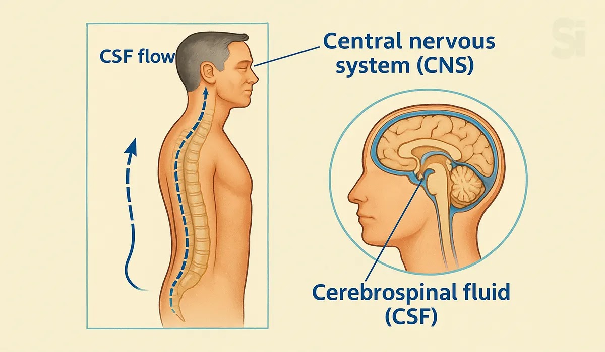
Diagram: Cerebrospinal fluid (CSF)
Functions of CSF
- It acts as a buffer to shock and protects the brain and spinal cord from damage due to body movement.
- It conveys nutritive material to the brain and spinal cord.
- It removes waste products from these structures.

Diagram: Functions of Cerebrospinal fluid diagram
FAQs
1. What is the basic function of the nervous system in the human body?
Answer: The nervous system controls body movements, senses, and thoughts. It
helps you react to your surroundings and keeps body functions running
properly.
2. Why should beginners learn about the nervous system?
Answer: Understanding the nervous system basics helps you know how your body
works. It is also a must-know topic for Medical Promotion Officer (MPO)
candidates.
3. What are the main parts of the human nervous system?
Answer: The main parts are the brain, spinal cord, and nerves. These parts
work together to send and receive messages in your body.
Conclusion
In this article, we explored The Nervous System and Its Functions in a clear
and simple way so that even beginners can easily follow along. If you are
aiming to build a career as a Pharmaceutical Sales Representative, this
foundational knowledge can give you an edge by helping you understand key
medical concepts with confidence.
Now you have a stronger idea of how the human nervous system works, and this
understanding will support you not only in your learning journey but also in
your job preparation. Keep expanding your knowledge step by step, because
the more you learn about medical science, the better equipped you’ll be for
success in the field.

Sarwar Hossain ✅
I am a travel enthusiast and sports-loving person. Education: BBA & MBA in Accounting.