Sensory organs of the human body | MPO training 12
As you train to be a Medical Promotion Officer (MPO), you need clear
information on the human senses. This article introduces the five key sensory
organs of the human body in simple words. It explains how eyes, ears, nose,
tongue, and skin help you see, hear, smell, taste, and feel. You will also
learn why knowing these organs is important for health communication.
The information is easy to read and friendly. By understanding our five
senses, you get a strong foundation for your MPO training and career. It gives
fun facts and useful tips that build your confidence in medical communication.
Table of contents: Sensory organs of the human body
Check out the table of contents below to learn everything you need to know
about sensory organs-
Sensory organs of the human body
Sensory organs of the human body are parts like the eyes, ears, nose,
tongue, and skin. They detect the five basic senses – sight, hearing, smell,
taste, and touch – by sensing light, sound, chemicals, and pressure. Each
organ sends information to your brain so you can recognize what you see,
hear, smell, taste, or feel. Knowing these organs helps MPO trainees explain
health topics clearly.
For example, understanding how eyes work can help you discuss vision care
with doctors. This basic knowledge forms a strong foundation for effective
communication in healthcare. They work together so you can respond to and
enjoy the world around you.
What are the sensory organs of the human body?
Our body has five main sensory organs that help us experience the world. The
five sensory organs are the eyes, ears, nose, tongue, and skin. Each one
helps you experience the world in different ways like seeing, hearing,
smelling, tasting, and touching.
These include:
| Sensory organs | Helps |
|---|---|
| Eyes: | For seeing light, color, and movement |
| Ears: | For hearing sounds and helping with balance |
| Nose: | For smelling different scents |
| Tongue: | For tasting flavors like sweet or sour |
| Skin: | For feeling touch, temperature, and pain |
Together, these organs send signals to our brain, helping us understand
and enjoy our surroundings.
Are there senses beyond the five main ones?
Yes. Besides the five main senses, our body has other ways to sense
things:
- Balance: Tiny parts in the inner ear let us know if we are upright or upside down.
- Body Awareness: Nerves in our muscles and joints tell the brain where our arms and legs are without looking.
- Temperature and Pain: Special nerves in our skin sense if something is hot, cold, or might hurt us.
These are not separate organs like eyes or ears, but they help us feel the
world. When people talk about sensory organs of the human body, they
usually mean the five main ones (eyes, ears, nose, tongue, and skin), but
our body has even more ways to sense things around us.
Special sense
The senses for which there are special end organs for the reception of
specific types of stimuli are called special senses.
Sensory Organs
Sensory organs have special receptors that allow us to smell, taste, see,
hear, and maintain equilibrium or balance.
The Information conveyed from these receptors to the central nervous
system is used to help maintain homeostasis. (Homeostasis: Normal body
balance)
The organs used for these functions are eye, ear, tongue and skin.
| Special sense | Sensory organ | Receptor |
|---|---|---|
| Vision | Eye | Rods and cones |
| Hearing | Ear | Hair cell of organ of corti |
| Olfaction or smell | Nose | Bipolar cell of olfactory mucosa |
| Taste | Tongue | Taste buds |
| Equilibrium | Ear | Vestibular apparatus |
Sensory Organs of Human Body Pie Chart
MPO Perspective: Common Medical Conditions by Sensory Organ
Common Sensory Organ Disorders (MPO Focus):
Sensory organ - Eye
More than half the sensory receptors in the human body are located in the
eyes, and a large part of the cerebral cortex is devoted to processing
visual information.
Our eyes work like natural cameras, helping us see the world. Light enters
through the front of the eye and reaches the back, where special cells
sense it. These cells detect light and color and send signals to our
brain. The brain then turns these signals into the pictures we see around
us. Because of our eyes and brain, we can recognize faces, read, watch
movies, and enjoy the beauty around us.
Accessory Structures of the Eye
The accessory structures of the eye include the eyelids, eyelashes,
eyebrows, the lacrimal (tearing) apparatus, and extrinsic eye muscles.
Anatomical parts of eye
There are three layers/tunics in the eye:
- Outer fibrous layer: It contains sclera and cornea
- Middle vascular layer: It consists of Iris, ciliary body, choroid. Together these three are called Uveal tract.
- Inner neural layer: Retina.
Anatomical parts of the eye are -
- Cornea
- Iris
- Pupil
- Lens
- Retina
- Macula
- Optic nerve
- Choroid and
- Vitreous
Cornea: Clear front window of the eye that transmits and focuses
light into the eye.
Iris: Colored part of the eye that helps regulate the amount
of light that enters.
Pupil: Dark aperture in the iris that determines how much
light is let into the eye.
Lens: Transparent structure inside the eye that focuses light
rays onto the retina.
Retina: Nerve layer that lines the back of the eye, senses
light, and creates electrical impulses that travel through the optic nerve
to the brain.
Macula: Small central area in the retina that contains special
light sensitive cells and allows us to see fine details clearly.
Optic nerve: Connects the eye to the brain and carries the
electrical impulses formed by the retina to the visual cortex of the
brain.
Vitreous: Clear, jelly-like substance that fills the middle of
the eye.
Physiology of Vision
Light waves from an object (such as a tree) enter the eye first through
the cornea, which is the clear dome at the front of the eye.
It is like a window that allows light to enter the eye.
The light then progresses through the pupil, the circular opening in the
center of the colored iris.
Fluctuations in the intensity of incoming light change the size of the
eye's pupil.
As the light entering the eye becomes brighter, the pupil will constrict
(get smaller), due to the pupillary light response.
As the entering light becomes dimmer, the pupil will dilate (get larger).
Initially, the light waves are bent or converged first by the cornea, and
then further by the crystalline lens (located immediately behind the iris
and the pupil), to a nodal point (N) located immediately behind the back
surface of the lens.
At that point, the image becomes reversed (turned backwards) and inverted
(turned upside-down).
The light continues through the vitreous humor, (the clear gel that makes
up about 80% of the eye's volume), and then, ideally, back to a clear
focus on the retina, behind the vitreous.
The small central area of the retina is the macula, which provides the
vision of any location in the retina.
In the retina, rods and cones initiate visual signals and relay them to
bipolar cells, which transmit the signals to ganglion cells.
The retina registers the tiny photons of light interacting with it.
Within the layers of the retina, light impulses are changed into
electrical signals.
Then they are sent through the optic nerve, along the visual pathway, to
the occipital cortex at the posterior (back) of the brain.
Here, the electrical signals are interpreted or "seen" by the brain as a
visual image.
Disorders of eye
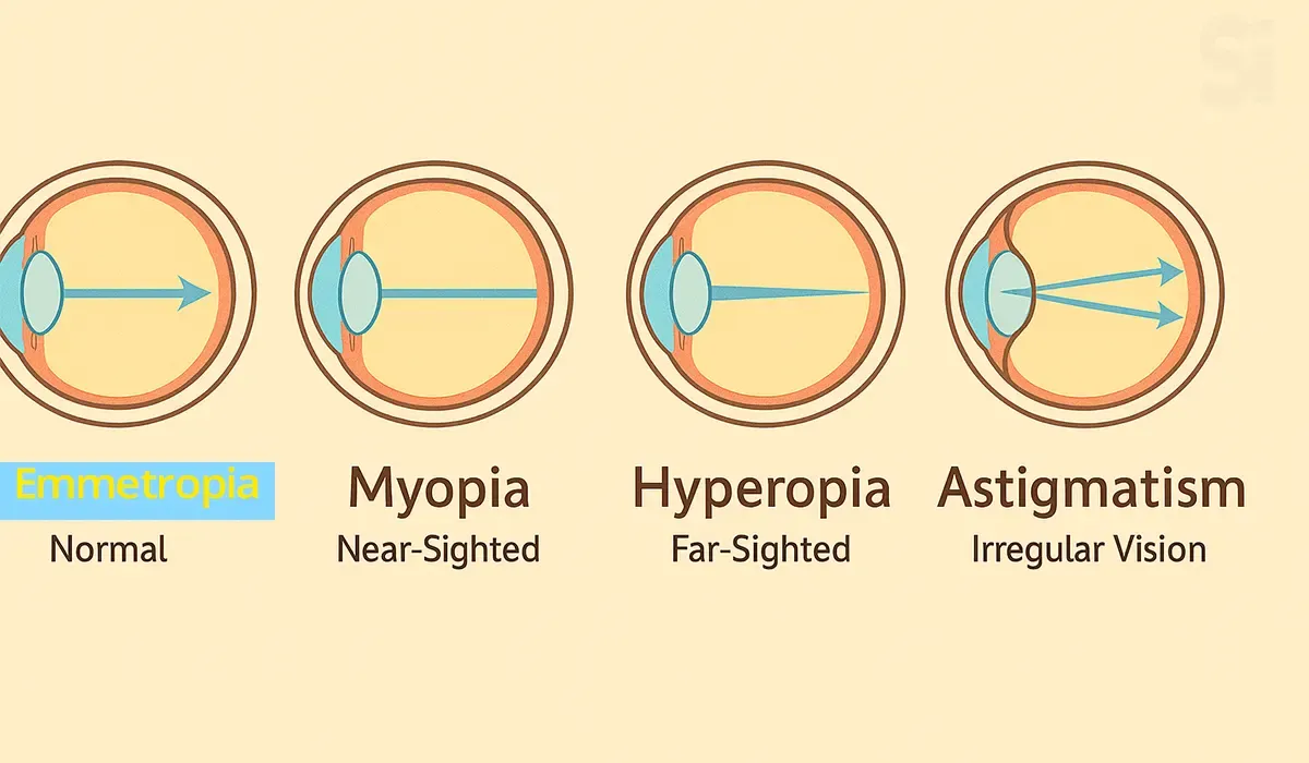
Myopia (Nearsightedness) : Near-sightedness is a common eye
condition in which faraway objects appear blurry.
Hyperopia (farsightedness) : Farsightedness means it's easy to see
things that are far away, but your closeup vision (near vision) is blurry.
Astigmatism : Astigmatism is a common vision problem caused by an
error in the shape of the cornea. With astigmatism, the lens of the eye or
the cornea, which is the front surface of the eye, has an irregular curve.
This can change the way light passes, or refracts, to your retina. This
causes blurry, fuzzy, or distorted vision.
Presbyopia : Presbyopia is an eye condition in which your eye
slowly loses the ability to focus quickly on objects that are close.
Cataract : A cataract is a clouding of the eye's natural
lens, which lies behind the iris and the pupil.
Glaucoma : Glaucoma is an eye disease that can damage your optic
nerve. The optic nerve supplies visual information to your brain from your
eyes. Glaucoma is usually the result of abnormally high pressure inside
your eye.
Corneal Ulcer : A corneal ulcer is an open sore that forms on the
cornea
Night blindness : Night blindness is also called "nyctalopia." It's
a type of vision impairment. People with night blindness experience poor
vision at night or in dimly lit environments.
Strabismus : Crossed eyes is also called strabismus, a condition in
which your eyes don't line up. If you have this condition, your eyes look
in different directions. And each eye will focus on a different object.
Blepharitis : Inflammation of the eyelids.
Conjunctivitis (Pink eye) : Inflammation or irritation of the
conjunctiva. It may be due to viral, bacterial or allergic inflammation.
Dry eye : A common condition that occurs when the eyes do not
produce enough tears to keep the eye moist and comfortable.
Keratitis : Inflammation of the cornea.
Sensory organ - Ear
The ear can transduce sound vibrations with small amplitudes into
electrical signals 1000 times faster than photoreceptors can respond to
light.
Our ears pick up sound waves and send them to the brain so we can hear.
Sound enters the ear and vibrates a drum-like part (the eardrum). These
vibrations travel through tiny parts in the ear and reach the inner ear,
where special cells turn them into signals. The brain receives these
signals and understands them as sounds like music, voices, or a car horn.
Besides receptors for sound waves, the ear also contains receptors for
equilibrium.
Anatomy of ear
The ear is divided into three main regions:
- The external ear, which collects sound waves and channels them inward.
- The middle ear, which conveys sound vibrations to the oval window.
- The internal ear, which houses the receptors for hearing and equilibrium.
External Ear
External ear is formed by two parts:
- Auricle or pinna
- External auditory meatus.
Middle Ear
Middle ear or tympanic cavity is a small, narrow, irregular, laterally
compressed chamber, situated within the temporal bone.
It is also known as tympanum. It is separated from external auditory
meatus by tympanic membrane.
Middle ear consists of the following structures:
1. Auditory ossicles
- Malleus
- Incus
- Stapes
2. Auditory muscles
3. Eustachian tube
Internal Ear
Internal ear or labyrinth is a membranous structure, enclosed by a bony
labyrinth in the petrous part of the temporal bone.
It consists of the sense organs of hearing and equilibrium. Sense organ
for hearing is the cochlea and the sense organ for equilibrium is the
vestibular apparatus.
Sensory Organ – Nose
The nose is a sensory organ that helps us to smell and also plays an
important role in breathing and filtering air.
Our nose has a special job: detecting different scents in the air. Inside
the nose are tiny cells that pick up scent particles from things we sniff.
These cells send a message to the brain, which figures out the scent. This
lets us enjoy nice smells (like flowers or baking bread) and notice
warnings (like smoke from a fire or spoiled food).
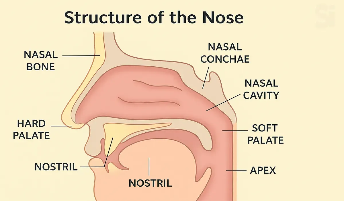
Structure of the Nose (Nose Anatomy in Simple Words)
- External Nose – The visible part outside the face.
- Nasal Cavity – The hollow space inside the nose.
- Nasal Septum – The divider that separates the left and right sides.
- Turbinates – Curved bones that help in filtering and warming air.
- Olfactory Region – The area responsible for smelling.
Functions of the Nose
| Function | Description |
|---|---|
| Smelling (Olfaction) | Detects smell using the olfactory nerve. |
| Breathing | Takes air in and out, helps respiration. |
| Air Filtration | Filters dust and germs using mucus and cilia. |
| Humidification | Adds moisture to the air. |
| Voice Resonance | Helps in clear and proper voice production. |
Smelling Pathway (Olfactory Pathway)
- Smell is detected by olfactory receptors.
- Signal goes to the olfactory bulb.
- Then it travels to the brain (olfactory cortex) to identify the smell.
Common Diseases Related to the Nose
| Disease | Explanation |
|---|---|
| Rhinitis | Inflammation of the nasal lining (can be allergic or due to infection). |
| Sinusitis | Infection or inflammation of the sinus cavities. |
| Nasal Polyps | Soft, painless growth inside the nose. |
| Anosmia | Loss of smell (can happen due to infection, trauma, or drug effect). |
| DNS (Deviated Nasal Septum) | The nasal divider is not in the center; can cause breathing problems. |
Sensory Organ – Tongue
The tongue is a muscular sensory organ inside the mouth. It plays a vital
role in tasting, speaking, swallowing, and chewing.
Our tongue is covered with many tiny bumps called taste buds. These taste
buds sense flavors like sweet, sour, salty, and bitter. When you eat
something, molecules from the food touch the taste buds on your tongue and
send signals to your brain. The brain figures out the flavor, so you know
if something is candy-sweet or lemony-sour. The tongue also helps us chew
food and speak clearly.
Structure of the Tongue (In Simple Terms)
1. Anterior 2/3 – Front portion; responsible for most taste sensation.
2. Posterior 1/3 – Back part; contains lymphoid tissue (part of tonsils).
3. Papillae – Small bumps on the tongue surface that contain taste buds:
- Fungiform
- Filiform
- Circumvallate
- Foliate
4. Muscles of Tongue – Help in movement during speaking, eating, etc.
5. Lingual Frenulum – A fold of tissue under the tongue that holds it in
place.
Functions of the Tongue
| Function | Description |
|---|---|
| Taste (Gustation) | Detects sweet, sour, salty, bitter, and umami tastes via taste buds. |
| Speech | Helps in pronunciation and clarity. |
| Swallowing (Deglutition) | Pushes food into the throat. |
| Mastication | Assists in chewing and mixing food with saliva. |
| Cleaning | Cleans mouth after eating. |
Taste Pathway (Simplified)
1. Taste buds detect the taste.
2. Signals are sent through cranial nerves:
- Facial Nerve (VII) – Anterior 2/3
- Glossopharyngeal Nerve (IX) – Posterior 1/3
- Vagus Nerve (X) – Base of tongue
3. Brain processes taste in the gustatory cortex.
Common Tongue Conditions
| Condition | Description |
|---|---|
| Glossitis | Inflammation of the tongue (due to infection or deficiency). |
| Geographic Tongue | Map-like appearance, usually harmless. |
| Oral Thrush | Fungal infection (white coating on tongue). |
| Taste Loss (Ageusia) | Can happen due to illness, drug reaction, or nerve damage. |
| Tongue-tie (Ankyloglossia) | Short frenulum limiting tongue movement. |
Sensory Organ – Skin
Skin is the largest sensory organ of the human body. It not only covers
and protects the body but also helps us feel touch, temperature, pressure,
and pain.
Our skin is the largest sensory organ in our body, full of nerve endings.
These nerves help us feel touch, temperature, and even pain. When
something touches your skin (like a soft blanket or a hot stove), the
nerve endings send signals to your brain. The brain then tells you what
you’re feeling – like hot or cold, soft or rough. This lets you enjoy
gentle touches (like a warm hug) and stay safe (by sensing if something is
too hot).
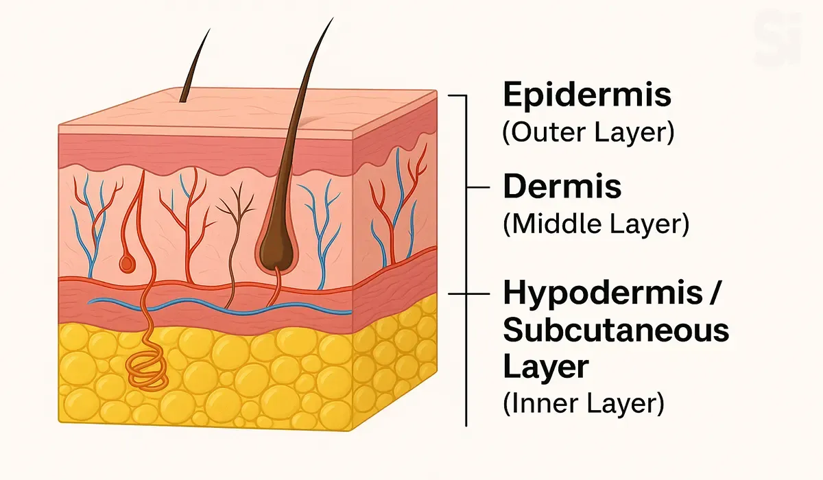
Structure of the Skin (In Simple Terms)
The skin has three main layers:
1. Epidermis (উপরের স্তর):
- Outermost layer.
- Contains keratin (protects against bacteria).
- Contains melanocytes (controls skin color).
- No blood vessels.
2. Dermis (মধ্য স্তর):
- Contains blood vessels, nerve endings, sweat glands, hair follicles, and touch receptors.
- Responsible for sensation and temperature regulation.
3. Hypodermis / Subcutaneous Layer (ভেতরের স্তর):
- Contains fat cells (adipose tissue).
- Acts as cushion and insulation.
Functions of the Skin
| Function | Description |
|---|---|
| Protection | Acts as a barrier against germs, chemicals, and UV rays. |
| Sensation | Detects touch, pain, heat, and pressure through sensory receptors. |
| Thermoregulation | Regulates body temperature through sweat glands and blood vessels. |
| Excretion | Removes waste (salt, urea) through sweat. |
| Vitamin D synthesis | Produces Vitamin D when exposed to sunlight. |
| Storage | Stores fat and water. |
Sensory Receptors in the Skin
| Receptor | Sensation |
|---|---|
| Meissner’s corpuscles | Light touch |
| Pacinian corpuscles | Deep pressure & vibration |
| Merkel discs | Steady pressure |
| Free nerve endings | Pain & temperature |
Common Skin Disorders
| Condition | Explanation |
|---|---|
| Dermatitis | Inflammation of skin (e.g., eczema, allergic reaction) |
| Psoriasis | Autoimmune skin condition with red, scaly patches |
| Fungal Infection | Ringworm, athlete’s foot, etc. |
| Acne | Inflammation of hair follicles and oil glands |
| Urticaria (Hives) | Itchy, allergic skin reaction |
To know more about skin and skin disease, first you have to know about the
integumentary system.
Equilibrium
Sense of Equilibrium
The sense of equilibrium consists of two parts:
- Static equilibrium
- Dynamic equilibrium
- The organs of static equilibrium help to maintain the position of the head when the head and body are still.
- The organs of dynamic equilibrium help to maintain balance when the head and body suddenly move and rotate.
Static Equilibrium
1. The organs of static equilibrium are located within the bony
vestibule of the inner ear, inside the utricle and saccule (expansions
of the membranous labyrinth).
2. A macula, consisting of hair cells and supporting cells, lies
inside the utricle and saccule.
3. The hair cells contact gelatinous material holding otoliths.
4. Gravity causes the gelatin and otoliths to shift, bending hair
cells and generating a nervous impulse.
5. Impulses travel to the brain via the vestibular branch of the
vestibulocochlear nerve, indicating the position of the head.
Dynamic Equilibrium
1. The three semicircular canals detect motion of the head, and they
aid in balancing the head and body during sudden movement.
2. The organs of dynamic equilibrium are called cristae ampullaris,
and are located in the ampulla of each semicircular canal of the inner
ear.
3. Hair cells extend into a dome-shaped gelatinous cupula. Rapid
turning of the head or body generates impulses as the cupula and hair
cells bend. Mechanoreceptors associated with the joints, and the
changes detected by the eyes also help maintain equilibrium.
How can we keep our sensory organs healthy?
We can protect the sensory organs of the human body by taking good care of
them in our daily lives.
Here are some simple tips:
- Eyes: Wear sunglasses on sunny days, and get regular eye check-ups.
- Ears: Turn down loud music and use earplugs in noisy places.
- Nose and Tongue: Don’t sniff or taste harmful chemicals, and keep your mouth and nose clean.
- Skin: Use sunscreen and wear gloves to protect your skin from sun and injury.
These steps help keep our eyes, ears, nose, tongue, and skin working well
so we can keep sensing the world.
How do sensory organs and our brain work together?
All the sensory organs of the human body send signals to our brain to help
us understand our surroundings. For example, our eyes send images of what
we see, and our ears send information about what we hear. The brain mixes
these signals together to create our full experience.
For instance, when you eat, your nose and tongue team up – your nose
smells the food and your tongue tastes it, and together they tell the
brain exactly what flavor you are enjoying. This teamwork helps you fully
enjoy events like watching and listening to a movie or smelling and
tasting a delicious meal.
Some brief notes
The human sense of hearing is attributed to the auditory system, which
uses the ear to collect, amplify, and transduce sound waves into
electrical impulses that allow the brain to perceive and localize sounds.
The ear can be divided into the outer ear, middle ear, and inner ear, each
of which has a specific function in the process of hearing.
The outer ear is responsible for the collection and amplification of
sound. The air filled middle ear transforms sound waves into vibrations,
protecting the inner ear from damage. The fluid-filled inner ear
transduces sound vibrations into neural signals that are sent to the brain
for processing.
The cochlea is the major sensory organ of hearing within the inner ear.
Hair cells within the cochlea perform the transduction of sound waves.
Humans are capable of estimating a sound's origin through a process called
sound localization, which relies on timing and intensity differences in
sound waves collected by each of our two ears.
FAQs
1. How do sensory organs help you in daily life?
Sensory organs help you stay aware of your surroundings. They protect you
from danger and help you enjoy life through sound, sight, smell, taste,
and touch.
2. How does the eye work as a sensory organ?
The eye captures light and sends signals to the brain so you can see. It
is a complex organ that helps you recognize color, shape, and distance.
3. What role does the ear play in the human body?
The ear helps you hear sounds and also controls balance. It sends sound
signals to the brain through tiny vibrations.
4. What is the function of the nose in the sensory system?
The nose detects smells. It also helps in breathing and filtering the air.
Smell signals go directly to the brain.
5. How does the tongue work as a sensory organ?
The tongue helps you taste different flavors like sweet, sour, salty,
bitter, and umami. It also helps in talking and swallowing.
6. What does the skin sense and how?
The skin is the largest sensory organ. It helps you feel temperature,
pressure, pain, and touch through nerve endings.
7. How can you protect your sensory organs?
You can protect your senses by avoiding loud noise, using sunglasses,
eating healthy foods, and avoiding harmful chemicals. Regular check-ups
are also helpful.
Conclusion
In conclusion, understanding the sensory organs of the human body is
important for anyone learning medical basics. This article showed you how
our eyes, ears, nose, tongue, and skin help us sense our world. You now
see why knowing these organs matters in health and MPO training.
Remember, this basic knowledge helps you talk confidently about medical
topics. Keep it in mind as you become an MPO. It lays a strong foundation
for your future career in medical communication. Value this knowledge as a
stepping stone to success in your healthcare communication career.


