Basics of the integumentary system | MPO training 11
The integumentary system is crucial for protecting the body and regulating
temperature. Understanding the basics of the integumentary system is essential
for anyone working in the medical field. As a Medical Promotion Officer (MPO),
having a clear grasp of this system will help you communicate better with
healthcare professionals.
Diagram: Integumentary system
This article provides a simple, easy-to-understand overview of how the skin,
hair, and nails work together to keep the body safe. Learn more about the
importance of the integumentary system and its role in health.
Table of contents: Basics of the integumentary system
Here's a quick look at what you'll learn from this article-
Basics of the integumentary system
The basics of the integumentary system involve understanding its main
components: the skin, hair, and nails. This system protects the body from
external harm, helps regulate temperature, and prevents dehydration. For a
Medical Promotion Officer (MPO), knowledge of this system is essential for
engaging with doctors and promoting health products effectively.
Knowing the functions of the integumentary system will improve communication
and enable you to explain its importance to healthcare professionals and
patients.
What is the integumentary system?
The integumentary system includes your skin, hair, nails, and various
glands. It acts as the body’s outer shield, protecting you from harm,
keeping your temperature in check, and letting you feel the world around
you.
The Integumentary System-
- Largest ongea.
- Composed of the skin, sweat and oil glands, hair, and nails.
- Accounts for 7% of the body's weight.
- Major role is protection from pathogens and dehydration.
- Varies in thickness from 1.5 to 4.0 mm.
- Composed of 3 distinct layers.
- Epidermis, Dermis, and hypodermis or superficial fascia.
Epidermis
- Outermost layer.
- Composed mostly of keratinized stratified squamous epithelium.
- Contains 4 distinct cell types and 4 to 5 distinct layers.
Diagram: Epidermis
Cell Types of the Epidermis
Keratinocytes:
Function: The main purpose of these keratin-producing cells is to
preserve against microbial, viral, fungal and parasitic invasion; to
protect against UV radiation; and to minimize heat, solute and water
loss.
Langerhans' cells:
- Arise from the bone marrow.
- Act as macrophages that activate the immune system.
Merkel cells:
- Present at the junction of the epidermis and dermis. Associated with sensory receptors.
Melanocytes:
- Synthesize melanin.
- Located at the deepest layer of the epidermis.
- The melanin is transferred to the keratocytes.
- Protects against UV damage.
Layers of the Epidermis
Diagram: Layers of the Epidermis
- Stratum basale
- Stratum spinosum
- Stratum granulosum
- Stratum lucidum
- Stratum corneum
The Dermis
- Made mostly of connective tissue.
- The hide of the human body.
- Richly innervated and vascularized.
- Contains the hair follicles, sweat glands, oil glands, lymphatic vessels, and many sensory receptors.
Diagram: Dermis
The Dermis layers
The Dermis Consists of 2 layers:
Papillary layer - areolar connective tissue, heavily
vascularized. Contains the dermal papillae, capillary loops, and
Meissner's corpuscles. In some areas these lie on top of the dermal
ridges. Cause the epidermal ridges that cause fingerprints.
Reticular layer - dense irregular connective tissue.
Skin Appendages
a. Sweat Glands-more than 2.5 million per person.
- Eccrine sweat glands-coil in the dermis, a duct leads to a pore at the skin's superficial surface.
- Apocrine sweat glands in the axillary and anogenital areas. Empty into hair follicles. Contains fatty substances and proteins. May cause body odor. Begin to function at puberty. May contain pheromones.
b. Ceruminous glands-secrete earwax.
c. Mammary glands-secrete milk.
d. produce ear.
e. Sebaceous Glands-oil glands. Found everywhere except the palms and
soles.
f. Secrete sebum. Usually secreted into hair follicles.
g. Whiteheads.
h. Blackheads.
i. Acne-staphylococcus.
j. Hair-covers the entire body except for the palms, soles, lips, nipples,
and parts of the genitalia.
k. Mostly dead keratinized cells.
Diagram: Skin Appendages
Hair
Parts of the hair:
1. Shaft
- Medulla
- Cortex
-
Cuticle
- Split ends
2. Root
3. Hair color
4. Hair follicle
- Hair bulb
- Root hair plexus
- Arrector pili-How and why is this important?
- Hair papilla
Diagram: Hair
Nails
- Modification of the epidermis.
- Composed of keratin.
- Composed of a free edge, body, and a root.
- Nail bed-epidermis under the nail.
- Nail matrix-growth occurs here.
- Lunula.
- Cuticle.
Diagram: Nails
Functions of the Integument
1. Temperature Regulation
- Sweat glands
- Vasodilation and vasoconstriction
2. Cutaneous Sensation
- Meissner's corpuscles
- Pacinian corpuscles
- Root hair plexuses
- Pain and heat/cold receptors
3. Metabolic Functions
- Vitamin D synthesis
4. Blood Reservoir
- Shunts more blood into the circulation when needed.
5. Excretion
6. Chemical/Biological/Physical barrier
Common Skin disorders in Bangladesh
- Scabies
- Psoriasis
- Eczema
- Dermatitis
- Fungal infection
- Candidiasis
- Tinea pedis/cruris/corporis etc.
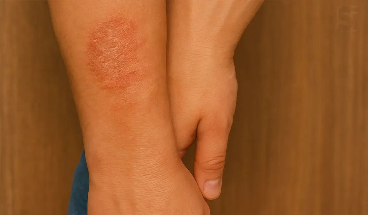
Psoriasis
Sign:
- Red patches of skin covered with thick, silvery scales
- Small scaling spots (commonly seen in children)
- Dry, cracked skin that may bleed or itch
- Itching, burning or soreness
- Thickened, pitted or ridged nails
- Swollen and stiff joints
Cause: genetics and the immune system.
Eczema
Sign:
- Rashes that commonly appear in the creases of the elbows or knees or the nape of the neck.
- Very dry skin on the affected areas.
- Rashes that are permanently.
- Itchy skin infections.
Cause: Experts aren't sure what exactly causes eczema. It may be
linked to your immune system's response to something irritating.
Fungal Infection
Sign:
- Irritation
- Scaly skin
- Redness
- Itching
- Swelling
- Blister
Cause/Organism: Trichophyton, Microsporum, Epidermo phyton,
Tinea manus, Tinea cruris, Tinea corpooris, Tinea capitis.
Tinea (pedis/cruris/corporis) infection
Sign: (Pedis)
- Skin which is red (rash-like and raw).
- Dry or moist and / or flaky.
- Cracked (fissuring) and scaly lesions (peeling) especially in the toe webbing and / or soles.
Sign: (Cruris)
- Looks like a ring. Due to irritation, the skin becomes red.
Sign: (Corporis)
- Scaly ring-shaped area, typically on the buttocks, trunk, arms and legs.
- A clear or scaly area inside the ring.
- Overlapping rings.
Scabies
Sign:
- Itching, often severe and usually worse at night.
- Thin, irregular burrow tracks made up of tiny blisters or bumps on your skin.
- The burrows or tracks typically appear in folds of skin. Though almost any part of the body may be involved.
Cause: Itch mite (Sarcoptes scabiei var. hominis).
Dermatitis
Sign:
- Usually beginning in infancy, this red, itchy rash usually occurs where the skin flexes inside the elbows, behind the knees and in front of the neck..
- The rash may leak fluid when scratched and crust over.
Cause: A gene variation, an immune system dysfunction, a skin
infection, exposure to food, airborne, or contact allergens, or a
combination of these.
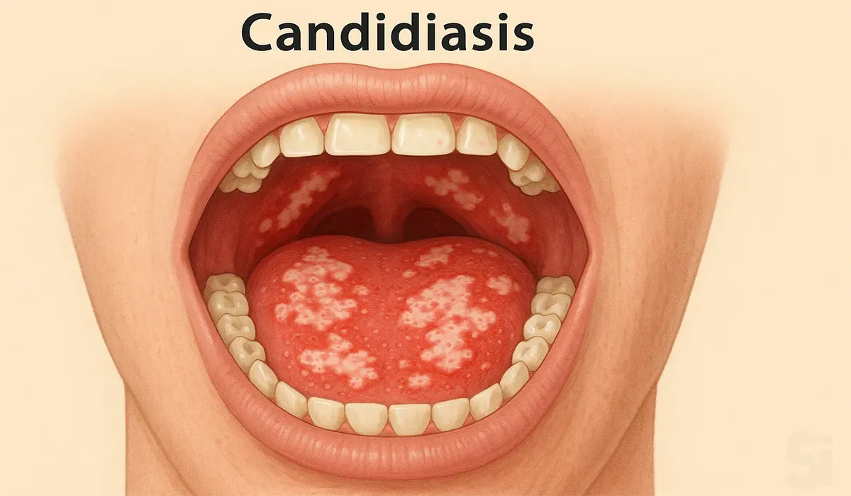
Candidiasis
Sign:
- A candida infection of the skin appears as a clearly defined patch of red, itchy skin, often leaking fluid.
- Oral thrush causes curd-like white patches inside the mouth, on the tongue and palate and around the lips.
Cause: Candida albicans.
Skin and soft tissue infections (SSTI)
These are bacterial infections of the skin, muscles, and connective
tissue such as ligaments and tendons.
Different types of SSTIs are:
- Impetigo
- Folliculitis
- Furuncles
- Carbuncles
- Erysipelas
- Cellulitis
- Pyoderma
- Acne etc.
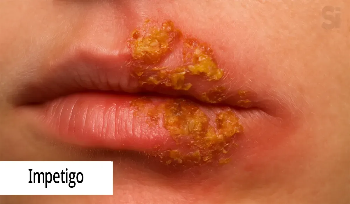
1. Impetigo: These are red sores, especially around the nose
and mouth. It's a skin infection caused by a bacteria, and it spreads
easily. It's most common in babies and young children, but adults can
get it too.
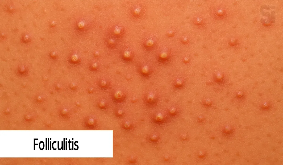
2.Folliculitis: Folliculitis is a common skin condition in
which hair follicles become inflamed. It's usually caused by a
bacterial or fungal infection.
3.Furuncles: This is an infection of the hair follicle that
goes into the deeper layers of skin. A small pocket of pus (abscess)
forms. It's also known as a boil. It is mostly caused by
staphylococcal infection.
4.Carbuncles: Carbuncle is a cluster of boils -painful,
pus-filled bumps that form a connected area of infection under the
skin.
5. Erysipelas: Erysipelas is an infection of the upper layers
of the skin (superficial). The most common cause is group A
streptococcal bacteria, especially Streptococcus pyogenes. Erysipelas
result in a fiery red rash with raised edges that can easily be
distinguished from the skin around it.
6.Cellulitis: Cellulitis is a common, potentially serious
bacterial skin infection. The affected skin appears swollen and red
and is typically painful and warm to the touch. Cellulitis usually
affects the skin on the lower legs, but it can occur in the face, arms
and other areas.
***Erysipelas and cellulitis are common infections of the skin.
Erysipelas is a superficial infection, affecting the upper layers of
the skin, while cellulitis affects the deeper tissues.
7. Pyoderma: Pyoderma gangrenosum is a rare condition that
causes large, painful sores ( to develop on your skin, most often on
your legs. The exact causes of pyoderma gangrenosum are unknown, but
it appears to be a disorder of the immune system.
8. Acne: Acne is a skin condition that occurs when your hair
follicles become clogged with oil and dead skin cells. It causes
whiteheads, blackheads or pimples.
FAQs
1) Why is the integumentary system so important?
This system is crucial because it guards against germs and physical
damage, helps regulate your body’s temperature, and prevents you
from losing too much water. It also enables you to sense touch,
pain, and temperature.
2) What are the main parts of the integumentary system?
The primary components are your skin, hair, nails, sweat glands, and
oil glands. Together, they help protect your body, keep you
comfortable, and aid in many other vital functions.
3) How does the skin protect the body?
Your skin serves as a strong barrier against harmful substances like
bacteria, chemicals, and physical injuries. It also keeps moisture
in and prevents dehydration, while helping regulate body
temperature.
4) What are the layers of the skin?
Skin is made up of three layers: the epidermis (outer layer), dermis
(middle layer), and hypodermis (deep layer). Each one has unique
functions, from protection to providing nutrients and helping with
temperature control.
5) How does the integumentary system help with temperature
regulation?
When you get hot, your sweat glands release sweat, cooling your body
down. If you’re cold, blood vessels in the skin constrict to keep
warmth inside, helping you maintain a stable temperature.
6) What role does hair play in the integumentary system?
Hair acts as a barrier, protecting your skin from the sun’s harmful
rays. It also helps trap heat when you’re cold, and it can alert you
to something lightly brushing against your skin.
7) How do nails contribute to the integumentary system?
Your nails are there to protect your fingertips and toes from
injury. They also play a role in your fine motor skills, like
grasping or scratching, making them an essential part of your
overall dexterity.
8) What’s the difference between sweat glands and sebaceous
glands?
Sweat glands produce sweat to cool your body down when it’s hot.
Sebaceous glands, on the other hand, secrete oil to keep your skin
and hair moisturized and protect them from drying out.
Conclusion
In conclusion, the basics of the integumentary system play a vital
role in maintaining overall health and well-being. For those working
as a Medical Promotion Officer (MPO), understanding this system is
key to promoting relevant medical products and educating healthcare
professionals. The integumentary system’s role in protection,
temperature regulation, and sensation is fundamental. With this
knowledge, you can effectively contribute to healthcare discussions
and be more proficient in your role as an MPO.


