Concept microbiology guide | MPO training 15
If you are preparing for a Medical Promotion Officer (MPO) job, this article
will help you understand microbiology in a simple way. The
Concept microbiology guide is designed for beginners like you who want
to learn the basics without any confusion.
It explains key ideas in easy language so you don’t feel lost. You will find
clear examples and real-life uses that match your MPO work. This guide is part
of a trusted MPO course that many already follow. Learn step-by-step with real
concepts, not just theory.
Table of contents: Concept microbiology guide
Check out the contents of this article or click where you want to go from
there-
Concept microbiology guide
The Concept microbiology guide helps you quickly understand important
microbiology terms that every Medical Promotion Officer (MPO) should know.
This article gives you clear, short explanations in easy language. You will
learn how these concepts apply in real MPO job roles.
It’s made for beginners, so you don’t need a science background. This guide
also includes simple examples and useful tips. If you want to grow your MPO
knowledge, this is a great place to start.
What is microbiology?
Microbiology is the study of tiny living organisms like bacteria, viruses,
fungi, and parasites that cannot be seen without a microscope.
Brief History of Microbiology
sir Antonie van Leeuwenhoek Von first observed and described bacteria in
1683. He made accurate descriptions of various types of bacteria. But the
significance was not realized. In 1857, Louis Pasteur established that the
fermentation is the result of microbial activity.
Microbiology - Microorganism & Pathogen
Microbiology : The science that deals with microorganisms.
Microorganism (Harmless) : An organism that can be seen only
through a microscope is called a microorganism. Microorganisms include
bacteria, virus, protozoa, algae and fungi.
Pathogen (Harmful) : A microorganism that causes the disease is
called a pathogen. Common pathogens are bacteria, viruses, protozoa and
fungi etc.
- Example: Streptococcus pneumoniae; which causes pneumonia. Salmonella typhi; which causes typhoid.
Microorganism & Pathogen
All pathogens are microorganisms but
all microorganisms are not pathogens.
Cause: Some microorganisms are harmless and even helpful. A
microorganism is only considered a pathogen if it causes disease.
Harmless viruses, bacteria, fungi, protozoa, and parasites are simply
called microorganisms.
- For example, Probiotics are live bacteria that are good for us, that balance our good and bad intestinal bacteria, and that aid in digestion of food and help with digestive problems, such as diarrhea and bellyache. Bacteria that are examples of probiotics are Lactobacillus and Bifidobacterium.
Pathology - Infection and Disease
Pathology : Pathology is the
scientific study of disease. Pathology is
concerned with the etiology (cause), pathogenesis
(development), and effects of disease.
Infection : The invasion and growth of the pathogen in the
body is called infection.
Disease : Disease is an abnormal state in which part
or all of the body is not properly adjusted or is incapable of
performing normal functions.
Bacteria - Virus and Fungus
| Bacteria | Virus | Fungus | Characteristics |
|---|---|---|---|
| Bacteria can only be seen under microscope | They are smaller than bacteria. They can only be seen under microscope | Most of them can be seen with naked eyes | Visibility |
|
Localized & Systemic (some extreme
cases) Body E.g. Sepsis
|
Systemic | Selectively localized & in extreme cases systemic | Infection Type |
| Generally tissue & organ | Any system | Generally specific tissue & organ | Affected Area |
| Antibiotics | Antivirals | Antifungal | Treatment |
| After completing Antibiotic Course | Usually 7-14 days | After completing Antifungal course | Recovery |
Bacteria
1. Glycocalyx
- The outermost layer of the cell.
- If it's dense and organized → called a capsule.
- If it's loose and unorganized → called a slime layer.
- Function: Protection from desiccation (drying), and helps evade host immune responses.
2. Cell Wall
- Provides shape and structural support to the bacterium.
- Protects from osmotic pressure and external damage.
3. Plasma Membrane
- A semipermeable membrane beneath the cell wall.
- Controls the movement of substances in and out of the cell.
4. Cytoplasm
- The internal fluid where various cell components float.
- Site of many metabolic reactions.
- Nucleoid (DNA)
- Region containing the bacterial chromosome (circular DNA).
- Controls cell activity and reproduction.
5. Plasmid
- Small circular DNA molecules separate from nuclei.
- Often carry genes for antibiotic resistance and other traits.
6. Ribosomes
- Site of protein synthesis.
- 70S type in prokaryotes.
7. Mesosome
- Infoldings of the plasma membrane.
- Involved in respiration and cell division.
8. Inclusion Bodies
- Reserve materials storage like nutrients, phosphate, etc.
9. Pili (singular: Pilus)
- Hair-like structures on the surface.
- Help in attachment and conjugation (gene transfer between bacteria).
10. Flagellum
1. Pilus: These are small hair-like structures on the surface.
They help the bacteria attach to surfaces or other cells.
2. Capsule: The outermost layer of the bacteria. It protects the
cell from drying out and helps the bacteria avoid the host's immune
system.
3. Cell Wall: A strong layer that gives shape to the bacteria and
protects it from bursting.
4. Plasma Membrane: This controls what goes in and out of the
cell. It helps maintain the internal environment.
5. Nucleoid (DNA): This is where the bacteria’s genetic material
(DNA) is found. It is not inside a nucleus like in human cells.
6. Cytoplasm: The jelly-like fluid inside the cell. All cellular
activities happen here.
7. Ribosomes: These are tiny structures that make proteins, which
are essential for the cell to function and grow.
8. Flagellum: A long tail-like structure that helps the bacteria
move from one place to another.
Classification of Bacteria
1. Shape
- Spirilla
- Bacilli
- Cocci
- Vibrio
2. Gram Stain
- Gram positive
- Gram negative
3. Oxygen Demand
- Aerobic
- Anaerobic
Shapes of Bacteria
* This image illustrates the "Shapes of Bacteria", showing the
different structural forms bacteria can take. It categorizes bacteria
based on their shape into five main types:
Cocci: These are spherical or round-shaped bacteria. They can
exist singly, in pairs (diplococci), in chains (streptococci), or in
clusters (staphylococci).
Bacillus: These are rod-shaped bacteria. They may occur singly
or in chains and some can form spores.
Spirilla: These are spiral-shaped bacteria that are rigid and
move using external flagella.
Vibrios: These bacteria are comma-shaped and slightly curved. A
well-known example is Vibrio cholerae, which causes cholera.
Spirochaetes: These are thin, flexible, spiral-shaped bacteria
that move in a corkscrew motion.
This diagram helps in understanding the morphological classification
of bacteria based on their shapes, which is important for
identification and diagnosis in microbiology.
* This image illustrates the shapes of bacteria, categorizing them
into three main types based on their morphology:
1. Spheres (Cocci) – Round-shaped bacteria:
- Diplococci (Streptococcus pneumoniae): Appear in pairs.
- Streptococci (Streptococcus pyogenes): Appear in chains.
- Tetrad: Groups of four cells arranged in a square.
- Staphylococci (Staphylococcus aureus): Grape-like clusters.
- Sarcina (Sarcina ventriculi): Cubic configuration of eight or more cells.
2. Rods (Bacilli) – Rod-shaped bacteria:
- Chain of Bacilli (Bacillus anthracis): Rods arranged in chains.
- Flagellate Rods (Salmonella typhi): Rods with flagella for movement.
- Spore-former (Clostridium botulinum): Rods that form spores under unfavorable conditions.
3. Spirals – Spiral-shaped bacteria:
- Vibrios (Vibrio cholerae): Comma-shaped.
- Spirilla (Helicobacter pylori): Rigid spiral-shaped bacteria.
- Spirochaetes (Treponema pallidum): Flexible spiral-shaped with more twists.
This classification helps in identifying bacteria and understanding
their behavior, which is crucial in microbiology and medical
diagnosis.
* This image illustrates the various shapes of bacteria, providing a
visual guide to help classify bacteria based on their morphology. It's
an educational diagram used in microbiology to identify and understand
different bacterial forms.
The diagram categorizes bacteria into distinct shapes and shows
examples of each type:
1. Spherical (Cocci):
- Staphylococcus aureus – Appears in grape-like clusters.
- Streptococcus pyogenes – Arranged in chains.
- Streptococcus pneumoniae – Appears in pairs (diplococci).
2. Rod-shaped (Bacilli):
- Bacillus cereus – Long, rod-shaped bacteria.
- Salmonella – Rod-shaped with flagella for mobility.
- Bordetella pertussis – Small, rod-shaped bacteria.
3. Comma-shaped (Vibrio):
- Vibrio cholerae – Curved or comma-like appearance.
4. Spiral-shaped (Spirilla and Spirochetes):
- Helicobacter pylori – Spiral-shaped with flagella.
- Treponema pallidum – Thin, tightly coiled (spirochete structure).
5. Other Notable Shapes:
- Klebsiella pneumoniae – Capsule-forming bacteria with irregular shapes.
- Corynebacterium diphtheriae – Club-shaped arrangement.
- Clostridium botulinum, Clostridium tetani – Spore-forming rod-shaped bacteria.
- Neisseria gonorrhoeae – Kidney bean-shaped diplococci.
This chart helps in understanding the morphological diversity of
bacteria. The shape of a bacterium can influence its mobility,
pathogenicity (ability to cause disease), and the method of treatment.
Gram Staining
Gram Staining Process:
This image illustrates the Gram staining technique, which is used in
microbiology to classify bacteria into
Gram-positive and Gram-negative
based on their cell wall structure.
1. Application of Crystal Violet (Primary Stain)
- The bacterial smear is first stained with crystal violet, a purple dye that enters all bacterial cells.
2. Application of Iodine (Mordant)
- Iodine is applied, which binds with the crystal violet and forms a complex that gets trapped in the cell wall.
3. Alcohol Wash (Decolorization)
- The slide is washed with alcohol.
- In Gram-positive bacteria, the thick peptidoglycan wall retains the violet stain.
- In Gram-negative bacteria, the thinner wall cannot retain the violet, so the stain is removed.
4. Application of Safranin (Counterstain)
Finally, safranin (a red/pink dye) is applied:
- Gram-positive bacteria remain purple.
- Gram-negative bacteria appear pink/red due to safranin.
Purpose: This method helps in identifying the type of
bacteria and guides appropriate antibiotic treatment.
Gram stain process
This image explains the Gram stain technique, which is a
fundamental microbiology method used to differentiate bacteria
into two main groups:
- Gram-positive and
- Gram-negative
based on their cell wall structure.
Step 1: Crystal Violet (Primary Stain)
- Purpose: This is the primary stain that is applied to all bacterial cells.
- Effect: All cells appear purple or blue initially.
Step 2: Iodine (Mordant)
- Purpose: Iodine binds with the crystal violet and forms a complex, making the dye less soluble and more firmly attached to the bacterial cell wall.
- Effect: All cells remain purple or blue.
Step 3: Alcohol (Decolorizer)
- Purpose: Alcohol washes the stain away from the thinner cell walls of Gram-negative bacteria.
-
Effect:
- Gram-positive cells: Remain purple or blue
- Gram-negative cells: Become colorless
Step 4: Safranin (Counterstain)
- Purpose: This is a secondary stain that colors the now-colorless Gram-negative cells.
-
Effect:
- Gram-positive cells: Stay purple or blue
- Gram-negative cells: Appear pink or red
Summary:
- Gram-positive bacteria → Appear purple or blue
- Gram-negative bacteria → Appear pink or red
This staining method is essential for identifying bacterial
species and guiding proper antibiotic treatment.
Morphological classification of Bacteria
A. Cocci
- Round in shape.
- Pairs is called Diplococcus.
- Chains is called Streptococcus.
-
Cluster is called Staphylococcus.
- Beta haemolytic streptococci Completely haemolyse RBC. E.g. Streptococcus pyogenes
- Alpha hemolytic streptococci Partially haemolyse. E.g. Streptococcus viridans
- Gamma hemolytic Streptococci No change in the RBC. E.g. Streptococcus faecalis
B. Bacillus
- Rod or cylindrical shaped bacteria are called bacilli.
- Pseudomonas aeruginosa
- Most bacilli are Gram negative
C. Vibrios
- They are comma shaped, curved rods.
- Vibrio cholerae: Produces cholera
D. Spirochaetes
- Spirochaetes Spiera means 'coil' and chaite means "hair".
- Treponema pallidum: Causes Syphilis
How are diseases caused by Bacteria ?
- Entrance (Portal of entry)
- Colonization (Adherence; Adhesion; Attachment)
- Multiplication with or without dissemination (spread)
- Penetration (Invasion)
- Signs and symptoms of disease (Morbidity; Mortality)
- Elimination and/or exit of pathogen (Carrier state may be established
Culprit Pathogen (Bacteria)
- Gram Positive Cocci
- Gram Negative Cocci
- Gram Positive Bacilli
- Gram Negative Bacilli
1. Gram Positive Cocci
- Staphylococcus aureus
- Staphylococcus epidermidis
- Streptococcus pyogenes
- Streptococcus viridans
- Streptococcus faecalis
- Streptococcus pneumoniae
- Peptostreptococcus
2. Gram Negative Cocci
- Neisseria gonorrhoeae
- Neisseria meningitidis
- Moraxella catarrhalis / Branhamell arrhalis
3. Gram Positive Bacilli
- Clostridium tetani
- Clostridium perfringens
- Corynebacterium diphtheriae
- Mycobacterium tuberculosis
- Clostridium botulinum
4. Gram Negative Bacilli
- Escherichia coli
- Proteus mirabilis
- Pseudomonas aeruginosa
- Salmonella typhi
- Salmonella paratyphi
- Haemophilus influenzae
- Haemophilus varias
- Klebsiella pneumoniae
- Bacteroides fragilis
- Shigella dysenteriae
- Yersinia enterocolitica
- Helicobacter pylori
- Bordetella pertussis
- Campylobacter jejuni
- Serratia marcescens
- Legionella pneumophila
- Vibrio cholerae
Extra:
1. Atypical Pathogen: Separate group — not classically
gram-stainable
- Mycoplasma pneumoniae
- Treponema pallidum
- Chlamydia trachomatis
Atypical bacteria cannot be classified as strictly
Gram-positive or Gram-negative because:
- They do not respond to classical Gram staining methods, or their staining is very weak.
- They have a different type of cell wall or lack a cell wall entirely (e.g., Mycoplasma).
- They typically have a unique mode of presentation, often being intracellular
Examples include:
- Mycoplasma pneumoniae → Lacks a cell wall (not Gram-stainable)
- Chlamydia trachomatis → Obligate intracellular
- Chlamydophila pneumoniae
- Legionella pneumophila → Difficult to stain with Gram stain; requires silver stain
- Coxiella burnetii
2. Anaerobic Pathogen (dual identity): Can be Gram-positive
or Gram-negative; extra label: anaerobic
- Bacteroides fragilis
- Fusobacterium
- Peptostreptococcus
Anaerobic bacteria are those that can survive — or even
thrive — in environments without oxygen. In fact, some may
die in the presence of oxygen.
These bacteria can be either Gram-positive or
Gram-negative, and include:
- Clostridium species (e.g., C. tetani, C. perfringens, C. botulinum) → Already listed under Gram-positive bacilli.
- Peptostreptococcus → Already listed under Gram-positive cocci.
- Bacteroides fragilis → Already listed under Gram-negative bacilli.
👉 Therefore, Anaerobic Pathogens represent a separate
classification based on oxygen requirements, but most of
them still fall under the categories of Gram-positive or
Gram-negative cocci/bacilli.
Virus
What is a virus?
A virus is an infectious agent of small size and
simple composition that can multiply only in
living cells of animals, plants, or bacteria. It has
no cell wall and no cell membrane.
Components of a Virus
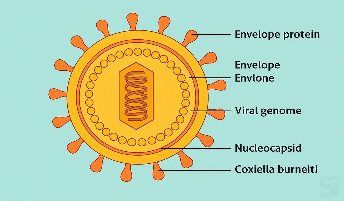
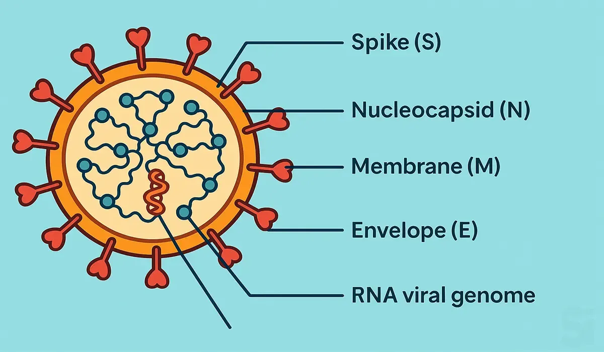
A fully assembled infectious virus is called a
virion.
Virions consist of two basic components:
- Nucleic Acid (single- or double-stranded RNA or DNA) and
- a Protein Coat.
The capsid is one kind of protein coat, which functions as a
shell to protect the viral genome and during infection
attaches the virion to specific receptors of the
host cell.
Some viruses contain an extra
lipid bilayer membrane surrounding the protein
capsid, which is called an envelope.
Classification of Virus Based on…
A. Classification Based on Nucleic Acid
- DNA Virus
- RNA Virus
(1) DNA Virus: A DNA virus is a virus that has a
genome made of deoxyribonucleic acid (DNA) that is
replicated by a DNA polymerase. Example: Herpes, Guap
smallpox, adenoviruses and papilloma viruses.
(2) RNA Virus: RNA virus is the virus that has
single-stranded as well as double-stranded RNA as its
genetic material.
Example: CoronaVirus, Hepatitis C Virus, Ebola Virus,
influenza Virus etc.
B. Classification Based on Capsid Symmetry or Position
- Coiled
- Spherical
(1) Helical Symmetry (Coiled): The helix (plural: helices) is a spiral shape that curves cylindrically around an
axis. In the case of a helical virus, the viral nucleic acid
coils into a helical shape and the capsid proteins wind
around the inside or outside of the nucleic acid, forming a
long tube or rod-like structure.
Example: Tobacco Mosaic Virus (TMV), Influenza virus
(2) Icosahedral Symmetry (Spherical-like): In
Icosahedral Capsid Structure, the genomes of icosahedral
viruses are packaged completely within an icosahedral capsid
that acts as a protein shell. Initially these viruses were
thought to be spherical.
Example: Poliovirus, Adenovirus.
C. Classification Based on the Presence of Envelope
- Enveloped
- Non-enveloped
(1) Enveloped: If the virus particle contains an
extra lipid bilayer membrane surrounding the protein capsid,
it's called an enveloped virus. Enveloped viruses are
typically less virulent than non-enveloped viruses. This is
because they don't always cause cell lysis during cell exit,
although cell death often follows as a consequence of virus
replication.
(1) Non-enveloped: Viruses are composed of two main
components:
- the viral genome (which can be RNA or DNA) and
- the virus-coded protein capsid
that surrounds the genome. If the virus particle contains
only these two elements, it is called a
non-enveloped virus. Non-enveloped viruses (also
known as naked viruses) are typically more virulent
than enveloped viruses. This is because they usually cause
host cell lysis.
Some Virus Related Terminologies
Nucleic Acid: A nucleic acid is an acidic,
chainlike biological macromolecule consisting of multiple
repeated units.
There are two types of nucleic acids:
- Deoxyribonucleic acid (DNA) and
- Ribonucleic acid (RNA).
The function of DNA is to store all of the genetic
information that to develop, function, and reproduce.
needs to develop function, and reproduce.
The primary biological role of RNA is to direct the
process of protein synthesis.
DNA Polymerase: DNA polymerase is an enzyme that is
responsible for forming new copies of DNA, in the form of
nucleic acid molecules.
RNA Polymerase: RNA Polymerase is an enzyme that
synthesizes RNA from a DNA template.
Transcription: The process by which DNA is copied
to RNA is called transcription.
Translation: The process by which RNA is used to
produce proteins is called translation.
Replication: Viral replication is the process by
which a virus makes copies of itself.
Fungi
What is Fungi?
Fungi are eukaryotic organisms that include microorganisms
such as yeasts, molds and mushrooms.
These organisms are classified under kingdom fungi.
Eukaryote: Any cell or organism that possesses a
clearly defined nucleus.
Structure of Fungi
The structure of fungi can be explained in the
following points:
- They can be either single-celled or multicellular organisms.
- Fungi consist of long thread-like structures known as hyphae.
- Almost all the fungi have a filamentous structure except the yeast cells.
- These hyphae together form a mesh-like structure called mycelium.
- Fungi possess a cell wall which is made up of chitin and polysaccharides.
- The cell wall comprises a protoplast, which is differentiated into other cell parts such as cell membrane, cytoplasm, cell organelles and nuclei.
- The nucleus is dense, clear with chromatin threads. The nucleus is surrounded by a nuclear membrane.
Characteristics of Fungi
The following are the important characteristics of
fungi:
- Fungi are eukaryotic organisms.
- They may be unicellular or filamentous.
- They reproduce by means of spores.
- Fungi lack chlorophyll and hence cannot perform photosynthesis.
- Fungi store their food in the form of starch.
- The nuclei of the fungi are very small.
- The fungi have no embryonic stage. They develop from spores.
- The mode of reproduction is sexual or asexual.
- Some fungi are parasitic and can infect the host.
Mold and Yeast
Mold: Molds are
eukaryotic multicellular microorganisms.
They have filamentous hyphae and airborne spores.
Molds decompose organic waste and are also used in
making antibiotics, cheese, etc. Few molds can also be
hazardous to health and can cause allergies,
headaches, itching, and respiratory problems.
Yeast: Yeast is a unicellular eukaryote.
Yeast is commonly found in fruits, animal skin,
vegetables, etc. It can convert carbohydrates to
alcohol and carbon dioxide through the process of
fermentation, which is an anaerobic process.
Yeast can also cause infections such as candidiasis in
humans. Saccharomyces cerevisiae is used in the baking
industry.
Fungal Cell Membrane
Ergosterol: Ergosterol is a sterol
found in the cell membranes of fungi and
protozoa. It acts to maintain cell membrane integrity,
similar to mammalian cholesterol.
Lanosterol: It is the compound from which all
animal and fungal steroids are derived.
Squalene: A precursor for the synthesis of all
plant and animal sterols (Steroid alcohol).
Ergosterol Synthesis
Squalene — (Squalene epoxidase)
---> Lanosterol
---------> Ergosterol synthesis (Essential
component of fungal cell membrane).
Note: Squalene epoxidase = Enzyme
Ergosterol synthesis is also catalyzed by a
CP450 enzyme called 14a-demethylase.
14a-Demethylase converts lanosterol to ergosterol in
fungi.
Concept microbiology guide FAQs
1) Why is microbiology important in the medical
field?
It helps us understand how infections start, how they
spread, and how to treat them with the right
medicines.
2) What are the main types of microorganisms?
The main types include bacteria, viruses, fungi,
protozoa, and algae.
3) What is the difference between bacteria and
viruses?
Bacteria are living cells that can survive on their
own, while viruses need a host (like the human body)
to live and multiply.
4) How do microorganisms cause disease?
Some microorganisms release toxins or invade body
tissues, which leads to infections and illness.
5) What is antibiotic resistance?
It means bacteria no longer respond to the antibiotics
used to kill them, making infections harder to treat.
6) What are common lab tests used in
microbiology?
Some common tests include culture tests, Gram
staining, and sensitivity tests to identify germs and
choose the right antibiotic.
7) What is a Gram-positive and Gram-negative
bacteria?
These terms describe how bacteria react to a special
stain. It helps doctors choose the right antibiotic.
8) How can you prevent infections caused by
microorganisms?
Good hygiene, clean water, safe food, vaccinations,
and using antibiotics properly help prevent
infections.
Conclusion
To become a successful Medical Promotion Officer
(MPO), you must understand the basics of microbiology
clearly. This Concept microbiology guide gives you the
right start with easy language and practical examples.
Whether you’re new or already preparing, this article
helps you learn faster. It focuses on real-life needs,
not just theory. You’ll feel more confident in your
MPO journey after reading it. This guide is your
helpful companion for smart learning.


